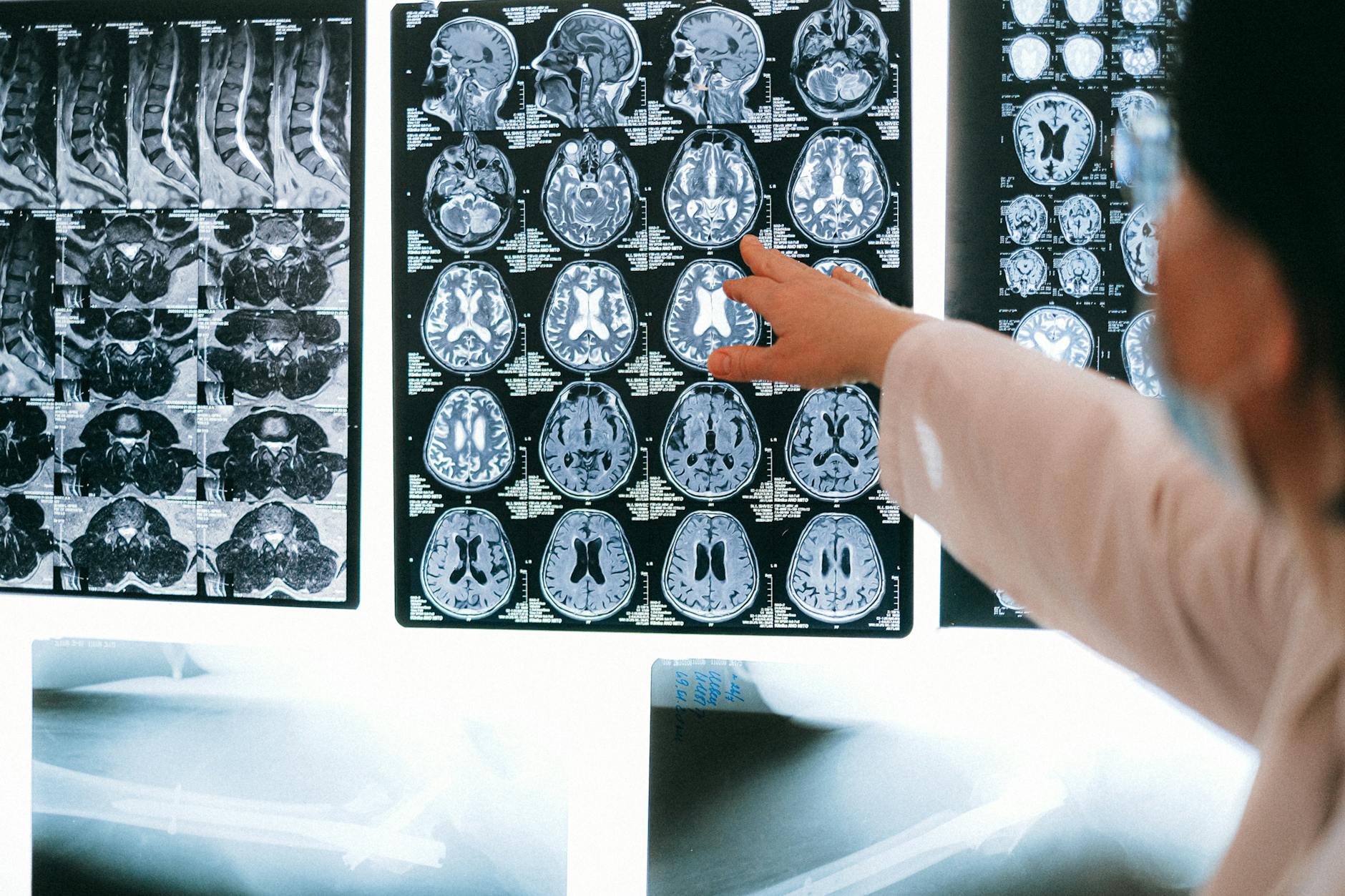
For Doctors in a Hurry
- Clinicians lack sensitive tools to detect subclinical neuronal damage in patients with chronic unilateral middle cerebral artery occlusion.
- The study utilized soma and neurite density imaging to evaluate 59 patients and 35 controls for gray matter microstructural changes.
- Patients exhibited significantly reduced soma and neurite signal fractions alongside increased extracellular space signal fractions across bilateral brain regions.
- The researchers concluded that right-sided middle cerebral artery occlusion causes more widespread bilateral damage than left-sided involvement.
- These findings allow for early identification of cognitive risks and support targeted interventions for patients with asymptomatic vascular disease.
The Hidden Cost of Chronic Cerebral Hypoperfusion
Chronic middle cerebral artery occlusion is often managed conservatively in asymptomatic patients, yet the long-term impact of persistent hypoperfusion on brain parenchyma remains a clinical blind spot. While conventional magnetic resonance imaging (MRI) is the gold standard for identifying acute infarction, it frequently fails to capture the subtle, progressive microstructural changes that precede overt tissue loss [1, 2]. Research into ischemic injury has increasingly focused on the complex interplay between axonal integrity, synaptic plasticity, and the neurovascular unit, which is the functional coupling between neurons, glia, and blood vessels [3, 4]. Furthermore, the role of neuroinflammation and glial responses, specifically the activation of resident immune cells like microglia to maintain homeostasis during chronic stress, suggests that "asymptomatic" may not mean "unaffected" [5, 6]. A recent study of 59 patients utilized soma and neurite density imaging (a diffusion MRI technique that quantifies the volume fractions of cell bodies and axons) to demonstrate that right-sided occlusion is associated with widespread bilateral microstructural damage involving the frontal, temporal, and limbic cortices [7]. These findings indicate that occult neuronal damage, characterized by significantly reduced soma and neurite signal fractions and increased extracellular space, may drive cognitive deficits even in the absence of acute stroke [7].
Quantifying Microstructural Decay via SANDI
The study cohort consisted of 59 patients with unilateral middle cerebral artery (MCA) occlusion who remained stroke-free despite chronic arterial blockage, alongside 35 control subjects for comparative analysis. To assess the integrity of the gray matter, all participants underwent a standardized imaging protocol that included three-dimensional T1-weighted imaging and a specialized diffusion magnetic resonance imaging technique known as soma and neurite density imaging (SANDI). This method allows clinicians to distinguish between different cellular compartments by modeling the diffusion of water molecules within the brain tissue, providing a more granular view of neuroanatomy than standard clinical sequences. By isolating the signal from different parts of the neuron, SANDI offers a window into cellular health before gross atrophy becomes visible on a standard T1 or T2 scan.
The researchers utilized model fitting of the SANDI data to generate high-resolution maps of four distinct microstructural parameters. These included the soma signal fraction (fSoma), which represents the volume fraction of cell bodies; the neurite signal fraction (fNeurite), reflecting the volume of axons and dendrites; the extracellular space signal fraction (fExtra), indicating the fluid environment outside the cells; and the apparent soma size (RSoma), which estimates the average radius of the neuronal cell bodies. To identify specific areas of degeneration, the team conducted a comprehensive voxel-based analysis (a method that examines every individual point in the three-dimensional image) employing a voxel-wise general linear model. This statistical framework allowed for a whole-brain comparison of RSoma, fSoma, fExtra, and fNeurite between the 59 patients with unilateral MCA occlusion and the 35 healthy controls, providing a detailed map of how chronic hypoperfusion alters the cellular landscape. This approach ensures that even small, localized clusters of neuronal loss are not overlooked by global averaging.
Hemispheric Asymmetry in Neurodegenerative Patterns
The spatial distribution of neuronal loss varied significantly depending on the side of the arterial blockage, revealing a distinct lack of symmetry in how the brain responds to chronic hypoperfusion. In patients with right-sided middle cerebral artery occlusion, the researchers observed widespread bilateral microstructural damage that extended far beyond the territory of the affected vessel. This extensive degeneration involved the frontal, parietal, temporal, occipital, and limbic cortices, as well as subcortical regions. Notably, this group also exhibited significant alterations in the hippocampus and parahippocampal gyrus, structures critical for memory and spatial navigation. The data indicate that damage is worse in right-sided middle cerebral artery occlusion cases compared to left-sided ones, suggesting that right-sided lesions may trigger more extensive network-wide failure across both hemispheres, perhaps due to the unique role of the right hemisphere in broad attentional and spatial networks.
Conversely, left-sided middle cerebral artery occlusion was characterized by left hemisphere-predominant damage, presenting a more localized pattern of neurodegeneration. In these patients, the microstructural changes were more restricted, showing more focal involvement of the frontal and parietal regions, along with the contralateral occipital regions. While the damage in left-sided cases was less extensive than in right-sided cases, the affected areas still encompass vital functional networks. Across the entire cohort of patients with identified abnormalities, the compromised brain regions involve the sensorimotor integration system, the language system, the limbic-emotional and memory system, subcortical gray matter, and the visual processing system. For the clinician, these findings suggest that even in the absence of an acute stroke, chronic occlusion of the middle cerebral artery leads to measurable decay in systems responsible for complex cognitive and motor functions, necessitating a more proactive diagnostic approach.
Clinical Implications for Cognitive and Functional Health
The primary goal of the study was to analyze gray matter damage in both the affected and contralateral hemispheres of patients with chronic arterial blockage. By mapping these microstructural changes, the researchers identified that the affected brain regions in unilateral middle cerebral artery occlusion encompass the sensorimotor integration system and the language system. Furthermore, the damage extends to the limbic-emotional and memory system, subcortical gray matter, and the visual processing system. These findings suggest that the clinical impact of chronic hypoperfusion is not limited to motor or sensory deficits but involves a broad spectrum of functional networks that govern complex behavior and communication. This broad involvement explains why patients may present with subtle executive dysfunction or mood changes that are often dismissed in the absence of visible infarcts.
The clinical risks appear most acute in patients with right-sided middle cerebral artery occlusion. In these cases, the microstructural damage frequently involved subcortical regions, the hippocampus, and the parahippocampal gyrus. Because these structures are essential for memory consolidation and spatial navigation, their involvement in right-sided middle cerebral artery occlusion patients often leads to cognitive deficits. This specific neuroanatomical pattern suggests that clinicians should prioritize cognitive screening in patients with right-sided lesions, even when they remain asymptomatic for traditional stroke symptoms. Identifying these deficits early may allow for more aggressive management of vascular risk factors to preserve remaining cognitive reserve.
At the cellular level, soma and neurite density imaging detected reduced fSoma and fNeurite (the signal fractions representing cell bodies and neuronal projections, respectively) alongside elevated fExtra (the signal fraction representing the extracellular space) in regions affected by the occlusion. Specifically, patients with unilateral middle cerebral artery occlusion demonstrate significantly reduced fSoma, significantly reduced fNeurite, and significantly increased fExtra in some regions of the bilateral hemispheres compared to healthy controls. These metrics provide a quantifiable marker of invisible neuronal loss, offering a basis for targeted clinical interventions and early risk stratification in patients who might otherwise be considered stable under conventional imaging protocols.
References
1. DiBella E, Sharma A, Richards L, Prabhakaran V, Majersik J, HashemizadehKolowri S. Beyond Diffusion Tensor MRI Methods for Improved Characterization of the Brain after Ischemic Stroke: A Review. American Journal of Neuroradiology. 2022. doi:10.3174/ajnr.a7414
2. Sokolowski JD, Soldozy S, Sharifi KA, et al. Preclinical models of middle cerebral artery occlusion: new imaging approaches to a classic technique. Frontiers in Neurology. 2023. doi:10.3389/fneur.2023.1170675
3. Li H, Li X, Xu Z, et al. Unbalanced Regulation of Sec22b and Ykt6 Blocks Autophagosome Axonal Retrograde Flux in Neuronal Ischemia–Reperfusion Injury. Journal of Neuroscience. 2022. doi:10.1523/jneurosci.2030-21.2022
4. Zhang D, Ruan J, Peng S, et al. Synaptic-like transmission between neural axons and arteriolar smooth muscle cells drives cerebral neurovascular coupling. Nature Neuroscience. 2024. doi:10.1038/s41593-023-01515-0
5. Kettenmann H, Hanisch U, Noda M, Verkhratsky A. Physiology of Microglia. Physiological Reviews. 2011. doi:10.1152/physrev.00011.2010
6. Amlerova Z, Chmelová M, Anděrová M, Vargová L. Reactive gliosis in traumatic brain injury: a comprehensive review. Frontiers in Cellular Neuroscience. 2024. doi:10.3389/fncel.2024.1335849
7. Liu Z, Wan X, Xiao Y. Invisible neuronal damage in stroke-free, asymptomatic chronic middle cerebral artery occlusion: evidence from soma and neurite density imaging.. European radiology. 2026. doi:10.1007/s00330-026-12495-7