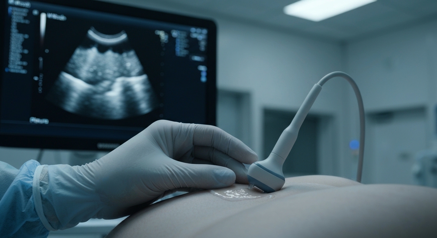
For Doctors in a Hurry
- Clinicians require more consistent, objective methods for interpreting lung ultrasound to diagnose pneumothorax at the bedside.
- The researchers developed an artificial intelligence model using 1,856 diverse ultrasound clips from patients, volunteers, and cadavers.
- The model achieved 100% sensitivity and 100% specificity, significantly reducing false positives compared to 11 experienced clinicians.
- The authors conclude that this explainable model matches the performance of an expert committee while minimizing diagnostic variability.
- This tool may serve as a reliable second reader to standardize bedside diagnosis and improve patient safety.
Standardizing the Sonographic Diagnosis of Pneumothorax
Bedside lung ultrasound has become a cornerstone of rapid assessment in critical care, offering superior sensitivity to chest radiography for detecting life-threatening conditions like pneumothorax [1, 2]. While its utility is well-established in neonatal care and emergency settings, the accuracy of the technique remains highly dependent on the skill and experience of the operator [3, 4]. Furthermore, the lack of transparency in automated diagnostic tools often hinders their integration into clinical workflows, as physicians require interpretable data to make high-stakes decisions [5]. A new study now investigates a method to standardize these interpretations through an explainable artificial intelligence framework.
Addressing Data Gaps with Diverse Training Sets
While lung ultrasound is essential for rapid, radiation-free bedside pneumothorax diagnosis, its clinical utility is frequently limited by significant variability in human interpretation. Current efforts to automate this process through artificial intelligence have faced several hurdles, including insufficiently large and diverse human datasets and inconsistent image acquisition across different clinical environments. Furthermore, existing artificial intelligence models often lack rigorous expert benchmarking and adequate clinical interpretability, leaving physicians without a clear understanding of how a machine reached a specific diagnostic conclusion. To address these limitations, the researchers developed an explainable soft-voting ensemble model, which is a machine learning architecture that combines predictions from multiple sub-models to reach a final consensus decision, much like a multi-expert consultation. The model was trained on a robust dataset of 1,856 diverse ultrasound clips, a sample size intended to capture a wide spectrum of pleural pathologies and artifacts. This training set was intentionally heterogeneous, incorporating imaging from critically ill patients, healthy volunteers, and tailored cadaver models to ensure the algorithm could handle the varied image quality and anatomical challenges encountered in real-world emergency and intensive care settings.
Benchmarking Against Expert Clinical Judgment
To evaluate the clinical utility of the ensemble model, the researchers conducted a rigorous benchmarking process against 11 experienced clinicians. This comparison utilized an independent, balanced test set, which is a separate group of ultrasound images not used during the training phase to ensure the results reflect real-world diagnostic accuracy. The statistical framework for this evaluation was comprehensive, incorporating measures of sensitivity, specificity, and inter-rater reliability (a metric used to quantify the degree of agreement between different observers). By comparing the AI directly to human experts, the study aimed to determine if the algorithm could provide a reliable second opinion in high-stakes clinical environments where diagnostic speed is critical. The results of the head-to-head comparison demonstrated that the AI ensemble model achieved 100% sensitivity (95% CI: 85.8% to 100.0%) and 100% specificity (95% CI: 85.8% to 100.0%). These figures indicate that the model correctly identified every instance of pneumothorax while also correctly ruling out the condition in every negative case within the test set. Notably, the AI model's sensitivity and specificity surpassed those of the expert clinicians, who showed greater variability in their assessments. This level of accuracy suggests that the AI ensemble matches the consensus-level performance of an expert committee, effectively functioning as a collective of specialists rather than a single observer. For the practicing physician, this means the tool could significantly reduce the risk of missed diagnoses or unnecessary interventions by providing a highly stable and accurate diagnostic baseline at the bedside.
Reducing False Positives in Challenging Imaging Modes
The clinical utility of lung ultrasound often depends on the imaging mode selected, and this study revealed that the diagnostic performance of experts significantly differed by ultrasound mode. While clinicians are trained to utilize both B-mode (real-time 2D imaging) and M-mode (motion mode, which captures movement over time along a single axis to visualize the 'seashore sign' or 'barcode sign'), the researchers found that expert clinicians showed notably lower specificity in M-mode imaging (p < 0.001). This statistical difference highlights a common clinical pitfall where the interpretation of motion artifacts in M-mode can lead to false-positive diagnoses of pneumothorax, potentially resulting in unnecessary and invasive procedures like chest tube insertions. In contrast, the AI consistently maintained perfect sensitivity across all conditions, ensuring that no true cases of pneumothorax were missed regardless of the imaging modality used. Beyond maintaining high sensitivity, the AI significantly reduced false positives compared to clinicians across all conditions, a finding that held true even in challenging diagnostic scenarios like subtle pleural motions. These subtle movements, often referred to as lung sliding or lung pulse, can be difficult for the human eye to distinguish from the absence of motion, particularly in critically ill patients with low tidal volumes. The ensemble model demonstrated excellent generalizability to both cadaveric and clinical cases, proving its robustness across different anatomical states and image qualities. For the practicing clinician, these results suggest that the AI tool can mitigate the diagnostic variability inherent in M-mode interpretation, providing a reliable safeguard against the overdiagnosis of pneumothorax in complex bedside environments.
Visualizing the Diagnostic Rationale
To address the common clinical concern regarding the opaque nature of artificial intelligence decision-making, the researchers prioritized model interpretability throughout the development process. They ensured transparency by using visualization through heatmaps (graphical representations where colors indicate areas of high diagnostic importance). These heatmaps allow the clinician to see exactly which anatomical regions the model is prioritizing, such as the pleural line or specific motion artifacts, when it identifies a pneumothorax or confirms the presence of lung sliding. This visual feedback transforms the AI from a black box into a collaborative tool that provides a clear rationale for its findings, mirroring the way a radiologist might point out specific features on a film during a consultation. The clinical relevance of these visual cues was confirmed through a rigorous verification process where the heatmaps generated by the AI were validated by expert clinicians. This step ensured that the model was focusing on the same physiological indicators used in manual interpretation rather than relying on irrelevant image noise or hardware artifacts. Because the model was trained on 1,856 diverse ultrasound clips and benchmarked against 11 experienced clinicians, its ability to highlight diagnostic landmarks provides a high level of confidence for the end user. By aligning the AI's focus with established sonographic criteria, the researchers created a system that supports, rather than replaces, the clinician's diagnostic workflow. For the practicing physician, the integration of this explainable model into the clinical environment offers a significant safeguard against medical error. The study findings demonstrate that the tool reduces diagnostic variability and false-positive diagnoses, which is particularly vital in high-pressure settings like the emergency department or intensive care unit where interpretation can be subjective. By acting as a reliable second reader, the AI helps prevent the unnecessary and invasive placement of chest tubes, which can occur when motion artifacts are misinterpreted as a pneumothorax. Ultimately, this ensemble model is designed to standardize clinical decisions at the bedside and substantially improve patient safety by providing a consistent, expert-level interpretation of lung ultrasound images.
References
1. Winkler MH, Touw H, Ven PMVD, Twisk JWR, Tuinman PR. Diagnostic Accuracy of Chest Radiograph, and When Concomitantly Studied Lung Ultrasound, in Critically Ill Patients With Respiratory Symptoms: A Systematic Review and Meta-Analysis. Critical Care Medicine. 2018. doi:10.1097/ccm.0000000000003129
2. Alrajab S, Youssef AM, Akkuş Nİ, Caldito G. Pleural ultrasonography versus chest radiography for the diagnosis of pneumothorax: review of the literature and meta-analysis. Critical Care. 2013. doi:10.1186/cc13016
3. Fei Q, Lin Y, Yuan T. Lung Ultrasound, a Better Choice for Neonatal Pneumothorax: A Systematic Review and Meta-analysis.. Ultrasound in medicine & biology. 2021. doi:10.1016/j.ultrasmedbio.2020.11.011
4. Chavez MA, Shams N, Ellington LE, et al. Lung ultrasound for the diagnosis of pneumonia in adults: a systematic review and meta-analysis. Respiratory Research. 2014. doi:10.1186/1465-9921-15-50
5. Chen H, Gómez C, Huang C, Unberath M. Explainable medical imaging AI needs human-centered design: guidelines and evidence from a systematic review. npj Digital Medicine. 2022. doi:10.1038/s41746-022-00699-2