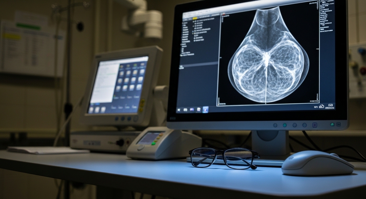
For Doctors in a Hurry
- Clinicians need to understand if artificial intelligence risk scores from prior mammograms predict future breast cancer characteristics.
- The study analyzed 1509 women who underwent breast biopsy in 2021 to evaluate these artificial intelligence risk scores.
- The area under the receiver operating characteristic curve for predicting biopsy-confirmed cancer was 0.62 with a 95% confidence interval of 0.59 to 0.65.
- The researchers concluded that these scores demonstrate modest discrimination and may capture imaging features associated with low-grade tumors.
- These findings suggest that artificial intelligence may identify subtle imaging patterns of malignancy one year before clinical detection.
Biological Correlates of Automated Mammographic Risk Assessment
Breast cancer remains the most frequently diagnosed malignancy and a leading cause of cancer death among women globally [1]. While mortality rates have declined by 40 percent since 1989 due to improved treatment and early detection, the incidence of hormone receptor positive and local stage disease continues to rise [2]. To address these trends, clinicians are increasingly exploring artificial intelligence as a tool to enhance screening accuracy and predict future malignancy risk [3]. Current evidence suggests that AI models can identify high risk patients with a degree of accuracy that often exceeds traditional clinical risk factors [3]. However, the specific pathological features that these algorithms detect in the years preceding a diagnosis remain poorly understood. A new study now investigates how these automated risk scores correlate with specific tumor characteristics to better understand the clinical relevance of early imaging signals.
The researchers conducted a retrospective study involving 1509 patients with a mean age of 58.56 ± 12.28 years who underwent breast biopsy following a screening mammogram in 2021 across four U.S. states. To evaluate the predictive capacity of automated assessment tools, the study utilized an FDA-approved artificial intelligence model to generate risk scores from screening mammograms performed in the year prior to the biopsy. This approach allowed the authors to investigate whether imaging features present 12 months before a clinical finding could signal the eventual development of malignancy. To evaluate the discriminative ability of these AI risk scores, the authors employed a receiver operating characteristic (ROC) analysis, which is a statistical method used to determine how effectively a diagnostic test can distinguish between two distinct groups, such as patients with and without cancer. Among the total cohort, 508 patients (33.7 percent) had biopsy-confirmed breast cancer. The analysis yielded an area under the ROC curve (AUC) of 0.62 (95% CI: 0.59–0.65) for the prior-year AI risk score in predicting biopsy-confirmed malignancy. This AUC value, which measures the probability that the model will rank a random positive case higher than a random negative one, indicates that the AI-generated risk scores demonstrated modest discrimination between biopsy-confirmed malignant and non-malignant cases. While the predictive power is not absolute, the findings suggest that the model identifies subtle imaging features that may precede clinical detection by at least one year, providing a potential window for earlier risk stratification.
Association Between AI Scores and Tumor Pathology
To further investigate the relationship between historical imaging data and specific pathological outcomes, the researchers utilized linear regression (a statistical method used to model the relationship between a dependent variable and one or more independent variables) to assess associations between prior-year risk scores and cancer characteristics among the subset of patients with confirmed malignancy. In a univariate analysis of these biopsy-positive cases, the study found that invasive lobular carcinoma (ILC) was associated with significantly higher prior-year AI risk scores compared to ductal carcinoma in situ (p = 0.009). Furthermore, the researchers observed a distinct correlation between the AI scores and the aggressiveness of the tumor; specifically, Grade 3 tumors had significantly lower scores than Grade 1 tumors (p = 0.016). The researchers then performed a multivariate analysis to determine if these associations remained robust after controlling for confounding factors. After adjusting for tumor grade, the association between invasive lobular carcinoma and AI risk scores was no longer significant (p = 0.136), which suggests that tumor grade may serve as a mediator in this relationship. In this adjusted model, Grade 3 tumors maintained a marginal association with lower risk scores (p = 0.068). These findings suggest that AI risk scores may reflect subtle imaging patterns of low-grade malignancy as early as one year before clinical detection. For the practicing clinician, this indicates that current AI models may be particularly sensitive to the slow-growing, architectural changes characteristic of low-grade tumors, potentially identifying evolving pathology before it meets traditional diagnostic thresholds. This sensitivity to low-grade features may eventually assist in tailoring screening intervals or supplemental imaging for patients whose historical scans show these subtle, high-risk signatures.
References
1. Jemal A, Bray F, Ferlay J, Ward E, Forman D. Global cancer statistics. CA A Cancer Journal for Clinicians. 2011. doi:10.3322/caac.20107
2. DeSantis C, Ma J, Gaudet MM, et al. Breast cancer statistics, 2019. CA A Cancer Journal for Clinicians. 2019. doi:10.3322/caac.21583
3. Schopf CM, Ramwala OA, Lowry KP, et al. Artificial Intelligence-Driven Mammography-Based Future Breast Cancer Risk Prediction: A Systematic Review.. Journal of the American College of Radiology : JACR. 2024. doi:10.1016/j.jacr.2023.10.018