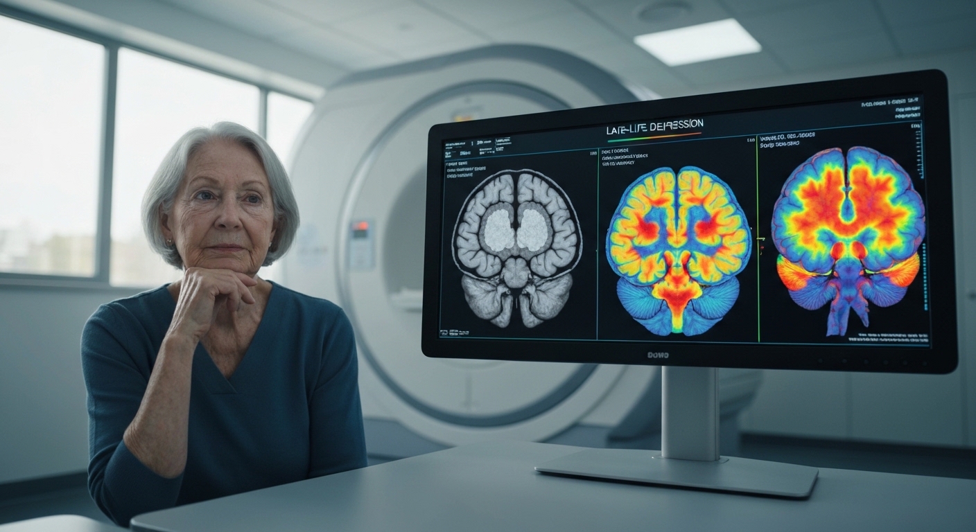
For Doctors in a Hurry
- Clinicians often struggle to differentiate Alzheimer's disease from late-life depression due to overlapping symptoms and unclear underlying biological markers.
- The researchers performed structural MRI scans on 33 patients with Alzheimer's disease, 38 with late-life depression, and 40 healthy controls.
- Patients with Alzheimer's disease showed extensive gray-matter atrophy in the hippocampus and cortex compared to the other two groups.
- The study concludes that these conditions share common biological pathways but follow distinct trajectories in brain structure and neurotransmitter profiles.
- These findings suggest that neurotransmitter-specific imaging may eventually assist in the differential diagnosis of these two complex clinical conditions.
The Diagnostic Dilemma of Cognitive Decline and Mood
Clinicians frequently encounter the diagnostic challenge of distinguishing between late-life depression and the early stages of Alzheimer’s disease, as both conditions often present with overlapping cognitive and neuropsychiatric symptoms [1, 2]. While late-life depression is a recognized risk factor for developing all-cause dementia, the biological transition from a primary mood disorder to neurodegeneration remains poorly understood [3, 4]. Current evidence-based guidelines emphasize the importance of early identification to implement risk-reduction strategies, yet neuropsychological assessments often fail to provide a definitive separation between these two pathologies [5, 6]. Recent advances in multimodal imaging and molecular mapping are now beginning to clarify the distinct structural and chemical signatures of these disorders [7]. A new study offers fresh insights into the divergent gray matter and molecular profiles that may allow for more precise clinical differentiation.
Mapping Structural Atrophy Across Patient Cohorts
The researchers recruited a clinical cohort consisting of 33 patients with Alzheimer's disease, 38 patients with late-life depression, and 40 age- and sex-matched healthy older adults who served as controls. To investigate the structural underpinnings of these conditions, all participants underwent high-resolution structural MRI using a 3.0 Tesla scanner. The team analyzed the resulting images using voxel-based morphometry (a computational technique that measures the volume of specific brain tissue types by comparing individual voxels to identify regional differences in gray matter density). This method allows clinicians to pinpoint exactly where brain tissue loss is occurring with high spatial precision. The analysis revealed that patients with Alzheimer's disease exhibited extensive gray-matter atrophy when compared to both the healthy control and late-life depression groups. When compared specifically to healthy controls, the atrophy in the Alzheimer's cohort predominantly involved the bilateral hippocampus, parahippocampal gyrus, cingulate cortex, and precuneus. These findings align with the established neurodegenerative progression of the disease, targeting regions critical for memory consolidation and self-referential processing. In a direct comparison between the two patient groups, the researchers found that the atrophy in Alzheimer's disease extended significantly further into the temporo-parieto-fronto-occipital association cortex than in late-life depression. Specific regions showing reduced volume in the Alzheimer's group compared to the depression group included the middle and inferior temporal gyrus, precuneus, insula, middle frontal gyrus, cingulate cortex, middle occipital gyrus, and medial superior frontal gyrus. Notably, the study found no significant gray-matter volume differences between the late-life depression group and the healthy controls, suggesting that the clinical symptoms of depression in older adults may not be driven by the same macrostructural neurodegenerative changes seen in Alzheimer's disease. This distinction is vital for the practicing physician, as it suggests that visible atrophy on a standard structural MRI in a symptomatic patient is highly suggestive of a primary neurodegenerative process rather than an isolated mood disorder.
Divergent Neurotransmitter and Synaptic Profiles
To move beyond macrostructural observations, the researchers employed the JuSpace toolbox to perform a cross-modal spatial correlation analysis (a computational method that links structural MRI findings with established molecular maps, effectively correlating regional gray matter changes with specific neurotransmitter profiles and cell surface markers). Through this analysis, the study identified that both the Alzheimer's disease and late-life depression groups exhibited abnormal associations with the 5-HT (serotonin) system. This shared serotonergic involvement may explain the overlapping affective symptoms often seen in both patient populations, such as anxiety and mood dysregulation, providing a biological basis for why both conditions sometimes respond to similar pharmacological interventions targeting serotonin pathways. Despite the shared serotonergic findings, the molecular signatures of the two conditions diverged significantly regarding other neurotransmitter systems and synaptic integrity. In patients with Alzheimer's disease, gray-matter reductions were negatively correlated with cholinergic and dopaminergic receptors, as well as the distribution of synaptic vesicle glycoprotein 2A (SV2A). Because SV2A is a reliable protein marker for synaptic density (the concentration of connections between neurons), its negative correlation with atrophy suggests that the structural loss in Alzheimer's disease is intrinsically tied to a widespread reduction in functional synapses. This molecular profile reinforces the clinical understanding of Alzheimer's as a primary neurodegenerative process characterized by the depletion of acetylcholine and dopamine signaling alongside the physical loss of synaptic connections. In contrast, the late-life depression group displayed a distinct neurochemical pattern where gray-matter reductions were positively correlated with NMDA (N-methyl-D-aspartate) receptors. Furthermore, the structural changes in the depression cohort were negatively correlated with the dopamine transporter (DAT) and cerebral blood flow. These findings suggest that the pathophysiology of late-life depression is more closely linked to glutamatergic signaling and vascular insufficiency rather than the broad synaptic loss seen in Alzheimer's. For the practicing physician, these results underscore that while the clinical presentation of these two disorders may appear similar, the underlying molecular drivers, particularly the roles of NMDA receptors and dopamine transporters, are markedly different, potentially necessitating distinct therapeutic approaches for cognitive and mood stabilization.
Cellular Phenotypes and Mitochondrial Dysfunction
The researchers extended their analysis to the cellular level to identify which specific neuron types were most associated with the observed structural changes. In the cohort of 33 patients with Alzheimer's disease, gray-matter reductions were negatively correlated with the distribution of projection neurons (the long-range excitatory cells responsible for communicating between different cortical regions) and somatostatin (SST) interneurons (a class of inhibitory cells that regulate dendritic inputs). Conversely, the study found that gray-matter reductions in Alzheimer's disease were positively correlated with parvalbumin (PVALB) interneurons. These PVALB cells are fast-spiking inhibitory neurons that provide perisomatic inhibition to principal cells. This divergent correlation suggests that while projection neurons and somatostatin-expressing cells are lost or diminished in areas of atrophy, parvalbumin-expressing cells may be relatively preserved or more densely concentrated in the remaining tissue of the Alzheimer's brain. The cellular profile of the 38 patients with late-life depression presented a different landscape of vulnerability. Unlike the Alzheimer's cohort, the late-life depression group showed a negative correlation between gray-matter volume and the distribution of PVALB neurons. This indicates that the structural integrity of the brain in depressed older adults is tied to the density of these specific inhibitory interneurons in a manner opposite to that seen in Alzheimer's disease. Additionally, late-life depression was negatively correlated with the granule-cell layer (a dense population of small neurons typically found in the dentate gyrus of the hippocampus and certain cortical layers). These findings highlight that the cellular underpinnings of volume loss in depression involve a distinct set of inhibitory and excitatory components compared to the neurodegenerative process of Alzheimer's disease. Despite these cellular differences, the study identified a shared metabolic vulnerability in both patient groups. Both the Alzheimer's disease and late-life depression groups showed weakened associations with mitochondrial complex I/IV function (the essential enzyme complexes within the electron transport chain responsible for cellular energy production). This commonality suggests that mitochondrial dysfunction and impaired energy metabolism represent a shared biological pathway underlying both conditions. Ultimately, the researchers concluded that Alzheimer's disease and late-life depression exhibit a partially overlapping yet markedly divergent profile across gray-matter atrophy patterns, neurotransmitter signatures, and cellular phenotypes. For the practicing physician, this confirms that while these two disorders may share certain metabolic stressors, they progress along distinct biological trajectories, requiring diagnostic and therapeutic strategies tailored to their unique molecular and cellular signatures.
References
1. Liu P, Xie Y, Meng X, Kang J. History and progress of hypotheses and clinical trials for Alzheimer’s disease. Signal Transduction and Targeted Therapy. 2019. doi:10.1038/s41392-019-0063-8
2. Invernizzi S, Bodart A, Lefebvre L, Loureiro IS. The role of semantic assessment in the differential diagnosis between late-life depression and Alzheimer's disease or amnestic mild cognitive impairment: systematic review and meta-analysis.. European journal of ageing. 2023. doi:10.1007/s10433-023-00780-z
3. Diniz BS, Butters MA, Albert SM, Dew MA, Reynolds CF. Late-life depression and risk of vascular dementia and Alzheimer's disease: systematic review and meta-analysis of community-based cohort studies. The British Journal of Psychiatry. 2013. doi:10.1192/bjp.bp.112.118307
4. Santabarbara J, Sevil-Perez A, Olaya B, Gracia-Garcia P, Lopez-Anton R. [Clinically relevant late-life depression as risk factor of dementia: a systematic review and meta-analysis of prospective cohort studies].. Revista de neurologia. 2019. doi:10.33588/rn.6812.2018398
5. Yu J, Xu W, Tan C, et al. Evidence-based prevention of Alzheimer's disease: systematic review and meta-analysis of 243 observational prospective studies and 153 randomised controlled trials. Journal of Neurology Neurosurgery & Psychiatry. 2020. doi:10.1136/jnnp-2019-321913
6. Barlet BD, Hauson AO, Pollard AA, et al. Neuropsychological Performance in Alzheimer's Disease versus Late-Life Depression: A Systematic Review and Meta-Analysis.. Archives of clinical neuropsychology : the official journal of the National Academy of Neuropsychologists. 2023. doi:10.1093/arclin/acad036
7. Marawi T, Ainsworth N, Zhukovsky P, et al. Brain-cognition relationships in late-life depression: a systematic review of structural magnetic resonance imaging studies. Translational Psychiatry. 2023. doi:10.1038/s41398-023-02584-2