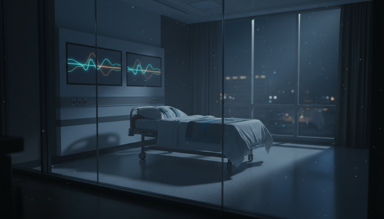
For Doctors in a Hurry
- Researchers investigated whether interictal breathing variability, or baseline respiratory fluctuations between seizures, predicts severe postictal hypoxemia.
- This prospective study analyzed video-electroencephalogram and respiratory data from 123 patients who experienced 152 generalized convulsive seizures.
- Awake long-term breathing variability significantly prolonged hypoxemia after seizures (mean ratio 1.06, 95% CI 1.01 to 1.13, p=0.04).
- The researchers concluded that greater respiratory variability between seizures correlates with prolonged and severe oxygen deprivation.
- Identifying this baseline respiratory dysregulation could serve as a clinical marker for sudden unexpected death in epilepsy.
The Silent Threat of Respiratory Dysregulation in Epilepsy
Sudden unexpected death in epilepsy (SUDEP) is the leading cause of epilepsy-related mortality in adults, yet predicting which patients are most vulnerable remains a persistent clinical challenge [1]. While the exact mechanisms are complex, evidence increasingly points to a cascade of centrally mediated cardiorespiratory failures, typically initiated by a generalized tonic-clonic seizure that triggers terminal apnea and subsequent cardiac arrest [2, 1, 3]. Research suggests that this vulnerability may stem from underlying defects in central respiratory control and chemoreception, leaving some individuals unable to recover from postictal oxygen depletion [4, 5]. Because these autonomic and respiratory deficits likely exist outside of the seizure event itself, investigators have begun searching for baseline physiological markers of respiratory instability. A new prospective study now investigates whether subtle irregularities in a patient's daily breathing patterns can predict the severity of oxygen desaturation immediately following a convulsive episode.
Capturing Respiratory Data in a Large Epilepsy Cohort
To investigate this potential link, researchers evaluated the relationship between interictal breathing variability (fluctuations in respiratory patterns between seizures) and the severity and duration of hypoxemia after generalized convulsive seizures. The prospective study collected comprehensive physiological data from people with epilepsy using continuous video-electroencephalogram (EEG), respiratory flow and effort monitoring, pulse oximetry (SpO2), and electrocardiogram (ECG). The research team initially enrolled 2,506 participants. Over the monitoring period, 257 participants experienced at least one generalized convulsive seizure. This subgroup included 141 females (approximately 54%) and had a mean age of 37.9 years. Because continuous physiological monitoring during violent motor seizures often introduces artifact interference, the final analysis was restricted to 152 seizure events in 123 participants who had evaluable respiratory data. Using this refined dataset, the investigators established specific clinical endpoints to quantify post-seizure respiratory failure. The first primary outcome was hypoxemia duration, defined as the length of time SpO2 was less than 90%. The second primary outcome was the severity of hypoxemia, defined as the SpO2 nadir (the lowest recorded oxygen saturation level following the seizure). Finally, the team assessed a secondary outcome, the occurrence of combined prolonged and pronounced hypoxemia, to identify patients experiencing the most extreme respiratory compromise.
Quantifying Baseline Breathing Irregularities
While a certain degree of fluctuation in the time between individual breaths is a normal physiological baseline, excessive irregularity can indicate underlying autonomic instability. The researchers hypothesized that increased variability in interbreath interval may suggest dysfunctional breathing control and may be associated with severe postictal hypoxemia. By identifying this baseline respiratory dysregulation, clinicians might be able to flag patients at the highest risk for dangerous oxygen drops following a convulsive seizure. To rigorously quantify these breathing patterns, the investigators analyzed continuous respiratory data collected outside of the seizure events during both asleep and awake periods. This allowed the team to assess respiratory control across different states of consciousness. To translate these continuous breathing patterns into actionable data, the researchers measured interictal interbreath interval variability using the coefficient of variation, root mean square of successive differences (RMSSD), and long-term (SD-2) variability from Poincaré plots. Each statistical tool captures a different dimension of respiratory instability. The coefficient of variation provides a standardized measure of overall dispersion in the breathing rate. The RMSSD evaluates short-term, breath-to-breath changes, highlighting immediate instability in the respiratory drive. Finally, the researchers utilized Poincaré plots (a geometric modeling technique that graphs each interbreath interval against the preceding one to visualize the predictability of the breathing cycle). From these plots, they extracted the SD-2 metric to quantify long-term variability trends, providing a comprehensive mathematical profile of each patient's baseline respiratory function.
Linking Interictal Breathing to Postictal Oxygen Drops
The researchers constructed multivariable models to evaluate how baseline physiological metrics influence the primary outcomes of hypoxemia duration and severity. In the multivariable model for hypoxemia duration, SpO2 nadir was significantly associated with the length of the desaturation event (mean ratio [MR] = 0.88, 95% CI 0.81-0.96, p = 0.002). Furthermore, baseline breathing irregularities during wakefulness predicted longer oxygen drops. Specifically, SD-2 of the awake interbreath interval was significantly associated with prolonged hypoxemia (MR = 1.06, 95% CI 1.01-1.13, p = 0.04). When examining the depth of oxygen desaturation, sleep-related breathing patterns proved critical. The RMSSD of the non-REM interbreath interval was the only variable significantly associated with hypoxemia severity (mean difference = -5.01, 95% CI -8.10 to -1.93, p = 0.002). This metric captures short-term, breath-to-breath instability during non-rapid eye movement sleep. Notably, the association between RMSSD of the non-REM interbreath interval and hypoxemia severity remained significant after controlling for duration of postictal generalized EEG suppression (a period of profound brain inactivity following a seizure), SD-2 of the awake interbreath interval, and body mass index. The investigators then evaluated the secondary outcome to identify patients who experienced the most severe respiratory compromise. Univariable analyses for combined prolonged and pronounced hypoxemia showed a significant association with SD-2 of the awake interbreath interval, reinforcing the link between long-term waking respiratory variability and severe post-seizure complications. Clinical seizure characteristics also played a role in this combined outcome. Univariable analyses showed a significant association with temporal lobe epilepsy. Additionally, specific intra-seizure respiratory and motor features were predictive, including a significant association with ictal central apnea (a cessation of breathing during the seizure itself) as well as a significant association with a shorter tonic phase duration.
Clinical Implications for SUDEP Risk Stratification
For clinicians managing patients with epilepsy, identifying who is most vulnerable to catastrophic outcomes remains a critical priority. The study demonstrates that measures of interictal respiratory variability are associated with severe and prolonged hypoxemia after generalized convulsive seizures. This relationship is clinically vital because severe hypoxemia after these seizures can trigger neural injury and is a potential biomarker for sudden unexpected death in epilepsy (SUDEP). By linking subtle, day-to-day fluctuations in breathing to the profound oxygen desaturation that follows a convulsive event, the researchers have identified a measurable physiological vulnerability. Ultimately, these findings point to a chronic autonomic deficit rather than just an acute reaction to a seizure. The authors conclude that increased interictal respiratory variability suggests baseline respiratory dysregulation in some people with epilepsy and may be a surrogate for SUDEP risk. For practicing physicians, monitoring these interictal breathing patterns could eventually provide a non-invasive tool to stratify patient risk. Identifying this baseline respiratory dysregulation may allow clinicians to implement targeted interventions, such as nocturnal oxygen monitoring or adjustments in antiseizure medications, for the patients most susceptible to fatal respiratory failure.
References
1. Jiang RY, Varughese RT, Kothare SV. Sudden Unexpected Death in Epilepsy: A Narrative Review of Mechanism, Risks, and Prevention. Journal of Clinical Medicine. 2025. doi:10.3390/jcm14103329
2. Ermongkonchai T, Kwok M, Prinsloo D, et al. Cardiac arrhythmias in sudden unexpected death in epilepsy: A systematic review. Epilepsy & Behavior. 2025. doi:10.1016/j.yebeh.2025.110582
3. Mir MY, Seh BA, Zahra S, Légrádi Á. The Crucial Interplay Between the Lungs, Brain, and Heart to Understand Epilepsy-Linked SUDEP: A Literature Review. Brain Sciences. 2025. doi:10.3390/brainsci15080809
4. Kim Y, Bravo E, Thirnbeck CK, et al. Severe peri-ictal respiratory dysfunction is common in Dravet syndrome. Journal of Clinical Investigation. 2018. doi:10.1172/jci94999
5. Dereli AS, Apaire A, Tahry RE. Sudden Unexpected Death in Epilepsy: Central Respiratory Chemoreception. International Journal of Molecular Sciences. 2025. doi:10.3390/ijms26041598