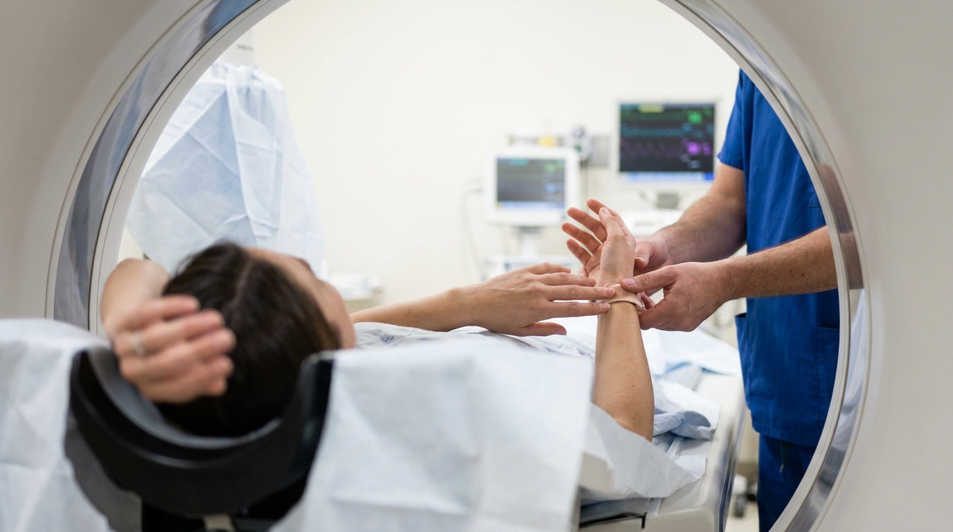
For Doctors in a Hurry
- Researchers investigated whether bilateral dynamic CT scanning with contralateral referencing improves the diagnosis of unilateral scapholunate ligament injuries.
- This retrospective multicenter study compared dynamic CT data from healthy volunteers and patients with arthroscopically confirmed unilateral scapholunate lesions.
- Total ligament injuries showed an increased scapholunate distance of 2.0 mm compared to the healthy contralateral wrist.
- The authors concluded that contralateral referencing in bilateral scanning effectively supports the diagnosis of total scapholunate ligament injuries.
- Using the patient's own healthy wrist as a reference eliminates the need for external datasets during diagnostic evaluations.
Refining the Diagnostic Workup for Scapholunate Instability
Diagnosing scapholunate interosseous ligament injuries remains a significant clinical challenge, as standard radiographs often fail to detect the dynamic instability that precedes carpal collapse [1, 2]. While magnetic resonance arthrography is frequently utilized for its high sensitivity to soft tissue tears, it cannot always characterize the functional impact of a lesion during active wrist movement [3]. Recent advancements in four-dimensional computed tomography have introduced the ability to visualize carpal kinematics in real time, offering a more detailed assessment of joint proximities during flexion and deviation [4, 5]. However, the lack of standardized protocols and the inherent variability in individual wrist anatomy have limited the widespread adoption of dynamic imaging in routine clinical practice [2, 6]. A recent study evaluates whether utilizing the contralateral wrist as an internal control can overcome these diagnostic hurdles and provide a more reliable baseline for identifying ligamentous incompetence.
Multicenter Cohort and Kinematic Methodology
This retrospective multicenter investigation was designed as a Level III diagnostic study to evaluate the efficacy of contralateral referencing, a technique that uses a patient's uninjured side as a baseline for comparison, in bilateral dynamic computed tomography scanning. While dynamic computed tomography enables three-dimensional assessment of wrist motion and is increasingly used to diagnose scapholunate ligament injuries, the lack of standardized reference values often complicates clinical interpretation. The researchers sought to determine if a patient's own healthy wrist could serve as a reliable internal control, thereby eliminating the need for external reference datasets that may not account for individual anatomical variation. The study combined dynamic computed tomography data from three distinct cohorts to provide a comprehensive comparison of carpal kinematics. These groups consisted of healthy individuals who underwent unilateral scans, healthy individuals who received bilateral scans, and a clinical group of patients with unilateral scapholunate lesions confirmed via arthroscopy who were scanned bilaterally. By comparing the healthy wrists of volunteers with the uninjured contralateral wrists of patients, the authors could assess whether injury to one side or general handedness significantly altered the baseline kinematics of the asymptomatic joint. To quantify the stability of the carpal bones during movement, the researchers utilized the scapholunate distance, a specialized measurement representing the width of the gap between the scaphoid and lunate bones. The scapholunate distance at five specific anatomical points was automatically extracted using computer-assisted techniques. These measurements were recorded while the subjects performed active wrist movements, specifically flexion-extension and radial-ulnar deviation. This approach allowed for a precise, objective analysis of how the scapholunate interval fluctuates throughout a functional range of motion, providing a more detailed diagnostic picture than static imaging to help clinicians better plan surgical or conservative interventions.
Addressing Protocol Variability and Natural Symmetry
A significant hurdle in the widespread adoption of dynamic computed tomography for wrist assessment is that protocol variability across different institutions limits the comparability and standardization of results. Currently, unilateral scanning (imaging only the symptomatic wrist) often requires clinicians to rely on external reference sets, which are databases of standardized wrist motion, to identify pathology. However, these external sets may not account for the unique anatomical nuances of an individual patient. To address this, the researchers assessed the influence of scan protocol variation on the scapholunate distance. The study found that differences in scan protocols led to measurable offsets in scapholunate distance of 0.5 mm, a margin that could potentially influence diagnostic accuracy when comparing results across different centers. These measured offsets in scapholunate distance were particularly evident during radial-ulnar deviation, the movement of the wrist toward the thumb or the pinky side. To determine if the patient's own anatomy could provide a more reliable baseline, the study also assessed wrist motion symmetry in healthy individuals. The researchers sought to confirm whether the uninjured side could serve as a valid internal control regardless of handedness. Their analysis revealed that minimal asymmetry was found between dominant and non-dominant wrists in healthy subjects. This high degree of natural symmetry suggests that the contralateral wrist provides a precise anatomical match for the patient. By utilizing bilateral scanning, clinicians can mitigate the 0.5 mm measurement discrepancies caused by institutional protocol variations. This approach allows for a personalized diagnostic comparison that eliminates the need for external reference datasets, ensuring that subtle changes in carpal mechanics are measured against the patient's own physiological norm.
Quantifying Scapholunate Dissociation in Total and Partial Tears
To validate the use of the uninjured side as a diagnostic benchmark, the study assessed the similarity between the wrists of healthy individuals and the contralateral wrists of patients. The researchers specifically evaluated contralateral referencing in patients with a unilateral scapholunate injury to determine if the presence of pathology on one side influenced the kinematics of the asymptomatic side. The analysis confirmed that contralateral healthy wrists of patients showed similar motion to that of healthy volunteers, suggesting that the uninjured wrist remains a stable and reliable internal control even in the presence of a contralateral ligamentous tear. The diagnostic utility of this bilateral approach is most evident when quantifying the degree of carpal dissociation. The researchers found that the magnitude of the gap between the scaphoid and lunate bones varied significantly based on the severity of the injury. In patients with total scapholunate injuries, an increased scapholunate distance of up to 2.0 mm was observed in the injured wrist compared to the contralateral side. This 2.0 mm threshold provides a clear metric for identifying complete ligamentous disruption during dynamic flexion-extension or radial-ulnar deviation. Detecting incomplete ligamentous damage requires even greater diagnostic sensitivity, as the deviations from the healthy baseline are far more subtle. The study found that partial scapholunate lesions showed smaller, localized asymmetries of approximately 0.3 to 0.4 mm when compared to the patient's healthy wrist. Because these measurements are smaller than the 0.5 mm offsets typically caused by variations in institutional scanning protocols, the use of the contralateral wrist as a reference is essential. This internal comparison allows clinicians to distinguish true pathological laxity from baseline anatomical variation or technical measurement noise, directly informing whether a patient might need surgical stabilization or conservative management.
Clinical Utility of Contralateral Referencing
The findings of this Level III diagnostic study indicate that contralateral referencing in bilateral dynamic computed tomography scanning is both possible and justified for supporting the diagnosis of unilateral total scapholunate injuries. By using the patient's own healthy wrist as an internal control, clinicians can accurately identify the pathological widening of the scapholunate interval. The researchers demonstrated that in cases of complete ligamentous disruption, the injured wrist exhibits an increased scapholunate distance of up to 2.0 mm compared to the asymptomatic side. This internal comparison is particularly valuable because it accounts for the patient's unique carpal morphology, providing a personalized baseline that is more precise than generalized population averages. For the practicing clinician, the primary benefit of this bilateral imaging strategy is that contralateral referencing removes the need for external reference datasets, which are standardized tables of normal wrist measurements derived from separate populations. Relying on external data can be problematic due to the 0.5 mm offsets in scapholunate distance measurements caused by variations in institutional scanning protocols, particularly during radial-ulnar deviation. By scanning both wrists simultaneously or in succession using the same protocol, these technical offsets are neutralized. This ensures that the observed differences, such as the 0.3 to 0.4 mm asymmetries seen in partial lesions or the larger 2.0 mm gaps in total tears, reflect true pathology rather than imaging artifacts. Based on the evidence that healthy individuals maintain high kinematic symmetry between their dominant and non-dominant wrists, the researchers suggest that bilateral dynamic scanning should be considered in the diagnostic workup for suspected carpal instability. This approach simplifies the diagnostic process by eliminating the search for age-matched or sex-matched normative data. Instead, the clinician can rely on the high degree of natural symmetry to confirm a unilateral injury, providing a clear and reproducible metric for surgical planning or conservative management transitions.
References
1. Dietrich TJ, Toms AP, Cerezal L, et al. Interdisciplinary consensus statements on imaging of scapholunate joint instability. European Radiology. 2021. doi:10.1007/s00330-021-08073-8
2. Goelz L, Kim S, Güthoff C, et al. ACTION trial: a prospective study on diagnostic Accuracy of 4D CT for diagnosing Instable ScaphOlunate DissociatioN. BMC Musculoskeletal Disorders. 2021. doi:10.1186/s12891-021-03946-x
3. Chang EY, Bencardino JT, French CN, et al. SSR white paper: guidelines for utilization and performance of direct MR arthrography. Skeletal Radiology. 2023. doi:10.1007/s00256-023-04420-6
4. Trentadue TP, Thoreson AR, Lopez C, et al. Detection of scapholunate interosseous ligament injury using dynamic computed tomography-derived arthrokinematics: A prospective clinical trial. Medical Engineering & Physics. 2024. doi:10.1016/j.medengphy.2024.104172
5. Demehri S, Baffour F, Klein JG, et al. Musculoskeletal CT Imaging: State-of-the-Art Advancements and Future Directions. Radiology. 2023. doi:10.1148/radiol.230344
6. Dobbe JGG, Roo MGAD, Visschers JC, Strackee SD, Streekstra GJ. Evaluation of a Quantitative Method for Carpal Motion Analysis Using Clinical 3-D and 4-D CT Protocols. IEEE Transactions on Medical Imaging. 2018. doi:10.1109/tmi.2018.2877503