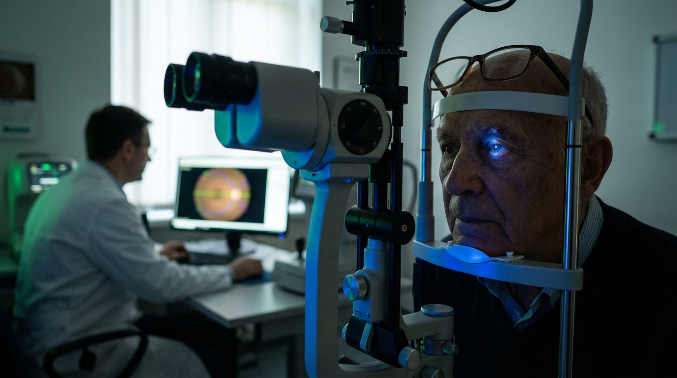
For Doctors in a Hurry
- Researchers investigated how the specific topographic distribution of geographic atrophy lesions impacts vision-related quality of life in patients with macular degeneration.
- This analysis included 856 participants aged 50 or older with bilateral geographic atrophy from two identical Phase III clinical trials.
- Geographic atrophy within the central 2.50-millimeter region independently associated with visual function scores (P < 0.001) with an R-squared of 0.11.
- The study concludes that vision-related quality of life depends more on lesion extent within the central 2.50-millimeter zone than peripheral areas.
- Clinicians should prioritize evaluating geographic atrophy extent within this central 2.50-millimeter region to better assess patient-reported visual impairment and functioning.
Topographic Determinants of Visual Function in Geographic Atrophy
Geographic atrophy represents a late, vision-threatening stage of age-related macular degeneration characterized by the irreversible loss of the retinal pigment epithelium, photoreceptors, and underlying choriocapillaris [1]. As the global prevalence of this condition increases, with estimates suggesting 10% to 20% of individuals over age 65 are affected, clinical focus has shifted toward preserving vision-related quality of life, which reflects a patient's self-reported ability to perform daily visual tasks and maintain independence [2, 3, 1]. While traditional management primarily involved antioxidant supplementation to reduce the risk of progression, the recent regulatory approval of intravitreal complement inhibitors, such as pegcetacoplan and avacincaptad pegol, has introduced active intervention [4, 5, 1]. Despite these therapeutic advances, clinicians often encounter a disconnect between the anatomical progression seen on imaging and the functional deficits reported by patients [6]. To bridge this gap, a recent analysis of 856 participants from the Chroma and Spectri trials identifies that geographic atrophy extent within the central 2.50-mm diameter region (P < 0.001) most significantly impacts self-reported visual function, regardless of whether the fovea is directly involved [7].
Quantifying Patient-Reported Outcomes in Phase III Trials
The researchers conducted a retrospective analysis of baseline data from 856 participants enrolled in the Chroma (NCT02247479) and Spectri (NCT02247531) trials. These two identically designed Phase III clinical trials investigated the efficacy of lampalizumab in patients 50 years of age or older who presented with bilateral geographic atrophy. By utilizing this large multicenter cohort, the study provided sufficient statistical power to evaluate how specific topographic patterns of retinal cell loss correlate with a patient's subjective visual experience.
To quantify vision-related quality of life, all participants completed the National Eye Institute Visual Function Questionnaire 25 (NEI VFQ-25) at the study's outset. The analysis focused on the visual functioning domain of the questionnaire, utilizing Rasch analysis (a psychometric model that converts simple ordinal survey rankings into linear interval scales) to refine the results. This statistical adjustment allows clinicians to treat survey responses as precise, continuous measurements, facilitating a more accurate comparison between subjective functional deficits and objective anatomical damage.
The anatomical assessment of these patients relied on advanced imaging to map the precise boundaries of retinal degeneration. Geographic atrophy lesions were automatically segmented using a combination of fundus autofluorescence and near-infrared reflectance images. While fundus autofluorescence identifies areas of metabolic stress and retinal pigment epithelium loss, near-infrared reflectance provides high-contrast structural detail of the atrophic boundaries. This dual-imaging approach allowed the researchers to calculate the minimum eye-level lesion extent across varying diameters, ranging from 0.25 mm to 6.00 mm relative to the fovea, to isolate the specific retinal regions that drive patient-reported disability.
The Critical 2.50-mm Central Zone
The researchers mapped the topographic distribution of lesions by measuring the extent of atrophy in central regions across varying diameters. These measurements began at a diameter of 0.25 mm and extended out to 6.00 mm, using precise 0.25-mm intervals relative to the fovea. To standardize the assessment across the cohort, the primary metric used was the minimum eye-level geographic atrophy extent in the region evaluated within an individual. This metric represents the smallest amount of atrophic tissue found within a specific zone for a given patient, providing a conservative estimate of retinal damage that accounts for the bilateral nature of the disease.
Statistical analysis demonstrated that the extent of atrophy across the entire macular area was linked to patient-reported outcomes. Specifically, the minimum geographic atrophy extent within all central regions between 0.25-mm and 6.00-mm diameters was significantly associated with NEI VFQ-VF person measures, which are the calibrated scores representing a patient's visual functioning (P ≤ 0.001 for all intervals). While the presence of atrophy anywhere in this 6.00-mm zone impacted quality of life, the strength of this association varied depending on the proximity to the foveal center.
The most clinically relevant finding emerged when comparing the predictive power of these different zones. The researchers found that the highest proportion of variance explained for vision-related quality of life occurred when evaluating the central 2.50-mm diameter region, yielding an R-squared value of 0.11. This R-squared value (a statistical measure representing the proportion of the variance for a dependent variable that is explained by an independent variable) indicates that atrophy within this specific 2.50-mm zone is a more potent driver of functional impairment than the total lesion area or atrophy in more peripheral regions. For the clinician, these data suggest that monitoring the expansion of lesions into this central 2.50-mm window may provide a more accurate prognosis for a patient's daily visual struggle than broader macular assessments.
Clinical Implications for Disease Monitoring
The researchers conducted a multivariable analysis to isolate which specific regions of the macula independently drive patient-reported outcomes. This statistical method, which accounts for multiple variables simultaneously to determine the unique contribution of each, demonstrated that the minimum geographic atrophy extent within the central 2.50-mm region was independently associated with NEI VFQ-VF person measures (P < 0.001). In contrast, the surrounding area, defined as the 2.50- to 6.00-mm annulus (the ring-shaped region outside the central zone), was not independently associated with these visual function scores (P = 0.541). These data indicate that once the damage within the central 2.50-mm zone is accounted for, the additional atrophy in the more peripheral macula does not significantly contribute to the patient's self-reported visual disability.
For the practicing ophthalmologist, these findings refine the structural parameters used to gauge a patient's functional prognosis. The study establishes that vision-related quality of life is most strongly associated with the minimum eye-level geographic atrophy extent within the central 2.50-mm region within an individual. This suggests that clinicians should prioritize the evaluation of the central 2.50-mm zone when monitoring disease progression and discussing lifestyle impacts with patients. Furthermore, the results indicate that evaluating geographic atrophy extent in this specific 2.50-mm region is more important than simply considering foveal involvement or total lesion size when seeking to understand the structural changes most closely linked to self-reported impairments in daily visual functioning.
References
1. Trincão-Marques T, Ayton L, Hickey DG, et al. Gene and cell therapy for age-related macular degeneration: A review.. Survey of ophthalmology. 2024. doi:10.1016/j.survophthal.2024.05.002
2. Vangsted A, Thinggaard BS, Nissen AHK, et al. Prevalence of geographic atrophy in Nordic countries and number of patients potentially eligible for intravitreal complement inhibitor treatment: A systematic review with meta‐analyses and forecasting study. Acta Ophthalmologica. 2023. doi:10.1111/aos.15768
3. Sharma A, Banait S. Treatment Paradigms in Age-Related Macular Degeneration: A Narrative Review Bridging Innovations in Dry and Wet AMD. Journal of Clinical and Diagnostic Research. 2026. doi:10.7860/jcdr/2026/81253.23402
4. Group AEDSR. A Randomized, Placebo-Controlled, Clinical Trial of High-Dose Supplementation With Vitamins C and E, Beta Carotene, and Zinc for Age-Related Macular Degeneration and Vision Loss. Archives of Ophthalmology. 2001. doi:10.1001/archopht.119.10.1417
5. Patel S, Lally DR, Hsu J, et al. Avacincaptad pegol for geographic atrophy secondary to age-related macular degeneration: 18-month findings from the GATHER1 trial. Eye. 2023. doi:10.1038/s41433-023-02497-w
6. Künzel SH, Broadbent E, Möller PT, et al. Association of Lesion Location and Functional Parameters with Vision-Related Quality of Life in Geographic Atrophy Secondary to Age-related Macular Degeneration.. Ophthalmology. Retina. 2024. doi:10.1016/j.oret.2024.01.025
7. Anegondi N, Lam D, Guymer RH, et al. Vision-Related Quality of Life in Geographic Atrophy: Association with Topographic Lesion Distribution.. Ophthalmology. 2026. doi:10.1016/j.ophtha.2026.04.019