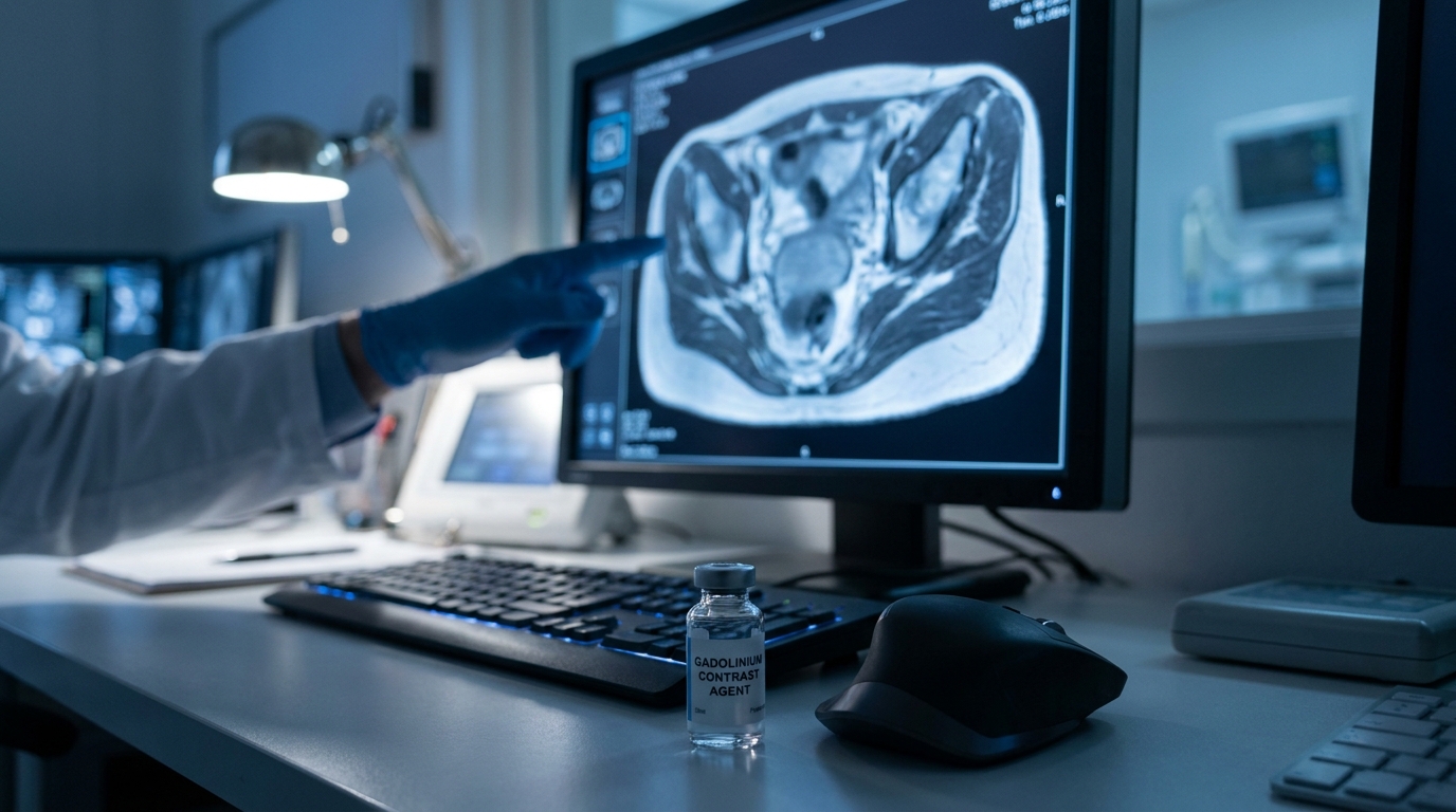
For Doctors in a Hurry
- Researchers investigated if adding contrast-enhanced sequences to MRI improves the identification of early-stage Tis-T1 rectal cancer compared to endorectal ultrasound.
- This study evaluated 136 patients with 138 confirmed rectal lesions using preoperative MRI and endorectal ultrasound to determine tumor staging.
- The combined MRI model achieved an AUC of 0.915, significantly exceeding the 0.806 AUC recorded for endorectal ultrasound (p = 0.003).
- The authors concluded that integrating the submucosal enhancing stripe into MRI protocols significantly increases diagnostic accuracy for early rectal lesions.
- These findings suggest that contrast-enhanced MRI provides a more reliable foundation for planning personalized organ-sparing treatments in early rectal cancer.
Refining the Preoperative Staging of Early Rectal Cancer
Colorectal cancer remains a leading cause of oncologic mortality in the United States, though survival rates improve dramatically when the disease is localized at the time of diagnosis [1, 2]. For patients with early-stage rectal lesions, the clinical priority is distinguishing between superficial tumors and those invading the muscularis propria to determine the feasibility of organ-sparing surgery [3, 4]. While endorectal ultrasound and pelvic magnetic resonance imaging are the standard modalities for local staging, their comparative accuracy in identifying the earliest T-stages has remained a subject of clinical debate [5, 6]. Current guidelines emphasize that precise pretreatment evaluation is the cornerstone of personalized surgical planning and long-term recurrence prevention [1, 7]. A recent study evaluates whether integrating specific contrast-enhanced features can improve the diagnostic performance of magnetic resonance imaging beyond these traditional benchmarks.
Evaluating Submucosal Integrity via Contrast Sequences
The researchers analyzed a cohort of 136 patients (mean age 60 ± 10 years, including 78 men) with pathologically confirmed Tis-T2 rectal cancer. Because all patients underwent curative resection between January 2020 and December 2023, the surgical pathology provided a definitive reference standard for the imaging findings. The analysis included a total of 138 rectal lesions, consisting of 82 Tis-T1 lesions and 56 T2 lesions. Before surgery, every patient received both a preoperative magnetic resonance imaging scan with contrast-enhanced sequences and an endorectal ultrasound. To refine the staging process, a radiologist evaluated tumor shape and the status of the muscularis propria (the integrity of the thick smooth muscle layer of the rectum) using standard T2-weighted images. A critical component of the investigation involved assessing the submucosal enhancing stripe, a specific feature visualized on contrast-enhanced sequences that represents the vascularized submucosal layer. By integrating these elements, the researchers constructed an MRI-based combined model utilizing tumor shape, muscularis propria status, and the submucosal enhancing stripe to identify stage Tis-T1 lesions. This integrated approach aimed to determine if adding contrast-enhanced features could provide a more granular view of the rectal wall layers than traditional imaging alone.
Superiority of Combined MRI Over Ultrasound
The study conducted a head-to-head comparison between the MRI-based combined model and endorectal ultrasound, which was performed independently by an endoscopist. To evaluate diagnostic performance in identifying stage Tis-T1 lesions, the researchers calculated the area under the receiver operating characteristic curve (AUC), a statistical metric where a value of 1.0 represents perfect diagnostic accuracy. Evaluating the status of the muscularis propria alone yielded an AUC of 0.762 (95% CI: 0.682-0.830), while assessing the submucosal enhancing stripe alone achieved an AUC of 0.861 (95% CI: 0.792-0.914). In comparison, the traditional endorectal ultrasound approach resulted in an AUC of 0.806 (95% CI: 0.730-0.868). This suggests that while ultrasound remains a standard clinical tool, its ability to differentiate early-stage lesions is limited compared to advanced imaging sequences. The most robust diagnostic performance was observed in the MRI-based combined model, which reached an AUC of 0.915 (95% CI: 0.856-0.956). This integrated approach significantly improved diagnostic accuracy over the common practice of assessing the muscularis propria alone, demonstrating an AUC difference of 0.154 (p < 0.001). Furthermore, the combined MRI model demonstrated higher diagnostic accuracy than endorectal ultrasound, showing a statistically significant AUC difference of 0.109 (p = 0.003). These findings indicate that adding contrast-enhanced features provides a more precise preoperative assessment than either ultrasound or standard T2-weighted imaging alone.
Clinical Implications for Organ-Sparing Surgery
The findings demonstrate that incorporating contrast-enhanced magnetic resonance imaging improves diagnostic accuracy for stage Tis-T1 rectal cancer compared to endorectal ultrasound, offering a highly precise tool for preoperative staging. By integrating the submucosal enhancing stripe with traditional T2-weighted features, the researchers achieved an overall diagnostic accuracy (AUC) of 0.915, which was statistically superior to the AUC of 0.806 provided by endorectal ultrasound (p = 0.003). For the practicing clinician, these results provide a more reliable basis for personalized, organ-sparing treatment planning. Accurately identifying Tis-T1 lesions is critical to avoid unnecessary radical resections, which carry higher morbidity, risk of permanent colostomy, and significant impacts on patient quality of life. By reducing the risk of overstaging T1 lesions as T2, the addition of contrast-enhanced imaging features provides a foundation for updating clinical practice guidelines. Ultimately, this integrated imaging approach allows gastroenterologists and colorectal surgeons to more confidently select appropriate candidates for less invasive surgical interventions, such as local excision or transanal endoscopic microsurgery.
References
1. Levin B, Lieberman DA, McFarland BH, et al. Screening and Surveillance for the Early Detection of Colorectal Cancer and Adenomatous Polyps, 2008: A Joint Guideline from the American Cancer Society, the US Multi-Society Task Force on Colorectal Cancer, and the American College of Radiology. CA A Cancer Journal for Clinicians. 2008. doi:10.3322/ca.2007.0018
2. Siegel RL, Kratzer TB, Giaquinto AN, Sung H, Jemal A. Cancer statistics, 2025. CA A Cancer Journal for Clinicians. 2025. doi:10.3322/caac.21871
3. Loft M, Pedersen M, Rahr H, Rafaelsen S. Can Ultrasound Elastography Discriminate between Rectal Adenoma and Cancer? A Systematic Review. Cancers. 2021. doi:10.3390/cancers13164158
4. Bonjer HJ, Deijen CL, Abis GA, et al. A Randomized Trial of Laparoscopic versus Open Surgery for Rectal Cancer. New England Journal of Medicine. 2015. doi:10.1056/nejmoa1414882
5. Luglio G, Pagano G, Tropeano FP, et al. Endorectal Ultrasonography and Pelvic Magnetic Resonance Imaging Show Similar Diagnostic Accuracy in Local Staging of Rectal Cancer: An Update Systematic Review and Meta-Analysis.. Diagnostics (Basel, Switzerland). 2021. doi:10.3390/diagnostics12010005
6. Fusco R, Petrillo M, Granata V, et al. Magnetic Resonance Imaging Evaluation in Neoadjuvant Therapy of Locally Advanced Rectal Cancer: A Systematic Review.. Radiology and oncology. 2017. doi:10.1515/raon-2017-0032
7. Singh HKSI, Lord A, Pawa N, Brown G. SRS68 - The prognostic impact of imaging detected Tumour deposits: a meta-analysis. British Journal of Surgery. 2026. doi:10.1093/bjs/znag018.078