- Clinicians need non-invasive myocardial characterization for patients with renal impairment who cannot tolerate gadolinium-based contrast agents during cardiac MRI.
- Researchers trained a model using 6,361 paired images from 1,189 subjects and validated it on 1,055 external test images.
- The model achieved AUC values of 0.906 and 0.895 for predicting enhancement probability while maintaining superior image quality (p < 0.05).
- The study concludes that contrast-free cine MRI effectively generates enhancement images comparable to the current clinical gold standard.
- This method provides a viable diagnostic alternative for patients with contrast contraindications, potentially increasing the accessibility of cardiac imaging.
Advancing Myocardial Tissue Characterization Without Gadolinium
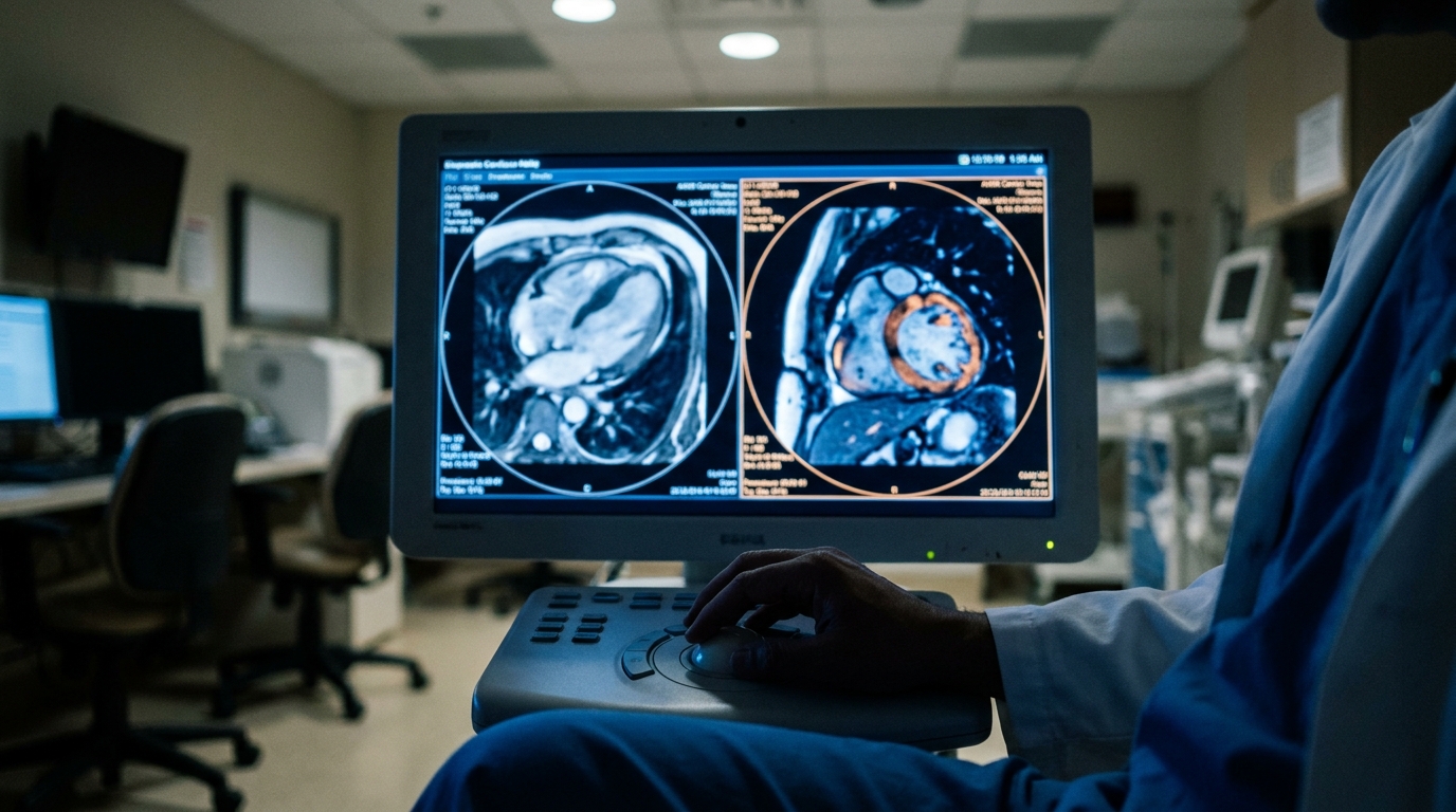
Late gadolinium enhancement (LGE) cardiac magnetic resonance imaging (MRI) remains the established clinical standard for non-invasive myocardial tissue characterization, providing essential data for the diagnosis and management of myocardial infarction [1][2]. In complex pathologies such as hypertrophic cardiomyopathy, LGE imaging is a critical component of risk stratification and diagnostic workups [3]. Beyond primary diagnosis, these sequences are increasingly utilized to evaluate tissue response and chronic scarring following therapeutic interventions like catheter ablation [4]. However, the requirement for gadolinium-based contrast agents presents a significant barrier for patients with advanced renal dysfunction or documented hypersensitivity. While various imaging strategies continue to emerge, the challenge remains to achieve high-resolution tissue characterization without the risks associated with exogenous contrast [4]. To address this clinical gap, researchers have evaluated a method to synthesize these diagnostic images using only standard, contrast-free sequences.
Synthesizing Enhancement from Standard Cine Sequences
The researchers developed Cine2LGE, a method based on a diffusion model (a type of generative artificial intelligence that creates high-resolution images by refining random noise into structured data). This computational framework simultaneously generates LGE-like images and estimates enhancement probability directly from contrast-free cine MRI, which are the standard motion sequences routinely used to assess cardiac function. By leveraging the underlying signal characteristics of non-contrast sequences, the model replicates the diagnostic information usually provided by gadolinium. For practicing cardiologists, this means potentially achieving accurate myocardial tissue characterization in patients for whom contrast is strictly contraindicated.
To ensure the tool remains robust across different clinical environments, the model was trained and evaluated on a multi-center, multi-manufacturer, and multi-disease dataset. This approach addresses a common limitation in medical imaging software by accounting for variations in scanner hardware and diverse cardiac pathologies. The training dataset comprised 6361 paired cine and LGE images from 1189 subjects, providing a large-scale foundation for the model to learn the complex mapping between standard cine sequences and the hyperintensity patterns seen in traditional LGE imaging.
Validation Across Internal and External Cohorts
To verify the clinical reliability of the Cine2LGE model, the researchers conducted a rigorous evaluation using both internal and external validation sets. The internal test dataset included 777 paired images from 129 subjects, providing an initial assessment of the model's ability to replicate enhancement patterns within the same clinical environment as the training data. To ensure the findings were not limited to a single institution or specific scanner settings, the model was also externally tested on an independent dataset consisting of 1055 paired images from 198 subjects. This external validation is critical for clinicians to determine if the tool can maintain its diagnostic performance across different patient populations and imaging protocols.
The assessment of image quality was performed by three independent, experienced radiographers who compared the synthetic images to traditional LGE scans. Beyond subjective quality, the researchers utilized a battery of statistical measures to confirm visualization consistency. They employed linear regression and correlation coefficient analysis to determine the strength of the relationship between the synthetic and real images. Furthermore, they used Bland-Altman plots (a statistical method used to evaluate the agreement between two different quantitative measurements by plotting the difference between them against their mean) to identify any systematic bias or outliers in the model's ability to represent myocardial tissue characteristics. These metrics ensure that the synthetic enhancement patterns are not only visually clear but also mathematically consistent with established gold-standard imaging.
Diagnostic Accuracy and Lesion Quantification
The clinical utility of Cine2LGE was primarily established through its ability to produce high-fidelity images that matched or exceeded the clarity of traditional contrast-enhanced scans. When evaluated by independent radiographers, the Cine2LGE model generated images with superior quality compared to conventional LGE (p < 0.05). This finding suggests that the diffusion-based synthesis process may mitigate some of the artifacts or noise often encountered in standard contrast protocols. Beyond visual clarity, the researchers focused on the quantitative reliability of the synthetic images for identifying myocardial damage. For the quantification of hyperintensity myocardial lesions (areas of dense scarring or acute injury), the Pearson correlation between Cine2LGE and traditional LGE was 0.732 in the internal set and 0.833 in the external set (p < 0.05). These coefficients indicate a strong linear relationship, suggesting that the synthetic images accurately reflect the extent of high-intensity tissue changes without the need for gadolinium.
The model also demonstrated precision in identifying more subtle tissue changes, such as intermediate-intensity myocardial lesions, which can represent diffuse fibrosis or border zones of an infarct. The Pearson correlation for intermediate-intensity lesion quantification was 0.759 in the internal set and 0.712 in the external set (p < 0.05), maintaining consistency across different patient cohorts. To evaluate the overall diagnostic accuracy of the system, the researchers used the area under the receiver operating characteristic curve (AUC, a statistical measure where a value of 1.0 represents perfect diagnostic discrimination). The enhancement probability prediction achieved an AUC of 0.906 on the internal test set and an AUC of 0.895 on the external test set. These high AUC values confirm that the model can reliably distinguish between enhanced and non-enhanced myocardium, providing clinicians with a robust tool for tissue characterization.
Clinical Utility for High-Risk Patient Populations
Late gadolinium enhancement cardiac MRI is currently the non-invasive gold standard for myocardial tissue characterization, yet its clinical application is frequently restricted. The reliance on gadolinium-based contrast agents limits LGE applicability in patients with renal impairment, who are at risk for nephrogenic systemic fibrosis, or those with documented contrast allergies. For these high-risk populations, the Cine2LGE model provides a software-based alternative by generating synthetic enhancement images directly from contrast-free cine MRI. This method utilizes a diffusion model (a generative artificial intelligence framework that synthesizes high-resolution images by refining random noise into structured data based on learned patterns) to estimate enhancement probability without exogenous agents. By removing the need for contrast, this approach may significantly expand the accessibility and clinical utility of cardiac MRI in routine practice, particularly for patients who were previously ineligible for standard tissue characterization protocols.
The clinical viability of this contrast-free method is supported by its performance across diverse patient cohorts. In a training dataset comprising 6361 paired cine and LGE images from 1189 subjects, the model learned to identify myocardial pathology with high precision. The researchers found that the enhancement probability prediction achieved an AUC of 0.906 on the internal test set and 0.895 on the external test set, where an AUC of 1.0 represents perfect diagnostic discrimination. Furthermore, the Cine2LGE model produced superior image quality compared to conventional LGE (p < 0.05), suggesting that the synthetic process may bypass common artifacts associated with contrast timing or patient breath-holding. These findings indicate that Cine2LGE is a viable alternative for patients who cannot undergo conventional LGE imaging, offering a reliable means of identifying myocardial lesions while eliminating the risks and costs associated with gadolinium administration.
Beyond simple detection, the model maintains quantitative rigor in assessing the extent of myocardial damage. Strong correlations were observed between Cine2LGE and traditional LGE in the quantification of hyperintensity lesions (Pearson correlation of 0.732 in the internal set and 0.833 in the external set; p < 0.05) and intermediate-intensity lesions (Pearson correlation of 0.759 in the internal set and 0.712 in the external set; p < 0.05). These metrics, which measure the linear relationship between the synthetic and real images, confirm that the model accurately reflects the volume of both dense scarring and diffuse fibrosis. For the practicing clinician, this means that Cine2LGE does not merely provide a visual approximation but offers a statistically robust tool for myocardial characterization that aligns closely with the established gold standard, potentially streamlining diagnostic workflows in cardiology departments.
References
1. Thygesen K, Alpert JS, Jaffe AS, et al. Fourth universal definition of myocardial infarction (2018). European Heart Journal. 2018. doi:10.1093/eurheartj/ehy462
2. Bøtker HE, Hausenloy DJ, Andreadou I, et al. Practical guidelines for rigor and reproducibility in preclinical and clinical studies on cardioprotection. Basic Research in Cardiology. 2018. doi:10.1007/s00395-018-0696-8
3. M EP, Elliott P, Anastasakis A, et al. 2014 ESC Guidelines on diagnosis and management of hypertrophic cardiomyopathy. European Heart Journal. 2014. doi:10.1093/eurheartj/ehu284
4. Hopman L, Pouderoijen NV, Mulder M, et al. Atrial Ablation Lesion Evaluation by Cardiac Magnetic Resonance: Review of Imaging Strategies and Histological Correlations.. JACC Clinical Electrophysiology. 2023. doi:10.1016/j.jacep.2023.08.013