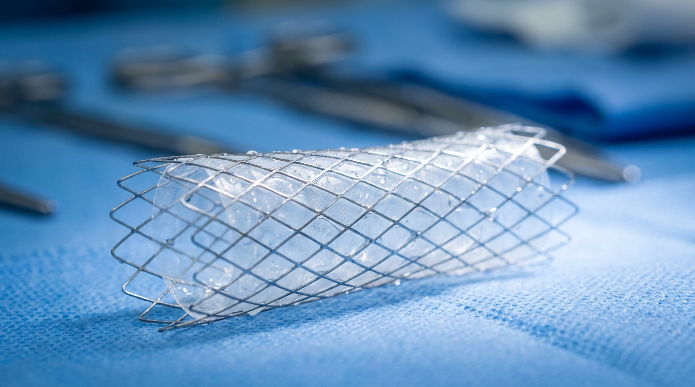
For Doctors in a Hurry
- Clinicians often struggle to manage life-threatening subclavian vein injuries due to the vessel's deep location and proximity to vital structures.
- The researchers evaluated the use of an 11 by 50 millimeter self-expanding stent-graft in a 60-year-old woman with iatrogenic injury.
- Emergency endovascular repair achieved immediate exclusion of the injury with no thrombosis or migration observed at 24 months of follow-up.
- The authors conclude that endovascular repair is a feasible and durable option for managing complex subclavian venous injuries.
- Physicians should prioritize rapid decision-making, appropriate anticoagulation, and rigorous postoperative surveillance when utilizing this endovascular technique for venous trauma.
Navigating the Challenges of Subclavian Venous Trauma
Iatrogenic injury to the great vessels of the upper thorax remains a rare but potentially catastrophic complication of head and neck surgery, often requiring immediate and decisive intervention. The subclavian vein is particularly vulnerable due to its deep anatomical position beneath the clavicle and its proximity to vital structures, making traditional open surgical repair technically demanding [1, 2]. While established guidelines emphasize the importance of rapid multidisciplinary intervention in vascular trauma, the optimal management of venous injuries in confined operative fields is still evolving [3, 4]. Current evidence suggests that standard compression or simple removal of offending catheters often results in poor outcomes, necessitating more robust reconstructive strategies [5]. Recent advances in endovascular technology have expanded the toolkit for managing complex vascular pathologies, yet clinical data on the long-term durability of venous stenting in this specific anatomical region remain sparse [6, 7]. A new case study involving the emergency deployment of an 11 x 50 mm Viabahn self-expanding stent-graft now provides critical longitudinal evidence, demonstrating full vessel patency at a 2-year follow-up following acute subclavian venous perforation [1].
Emergency Intervention for Iatrogenic Perforation
The clinical scenario involved a 60-year-old woman undergoing a lateral cervical lymph node dissection as part of her treatment for invasive thyroid carcinoma, a procedure that requires meticulous dissection near major vascular structures. During the surgical procedure, the patient sustained an iatrogenic injury of the left subclavian vein, a complication that immediately escalated into a life-threatening event. Surgeons initially attempted to achieve open hemostasis, but these efforts were unsuccessful. The failure of traditional surgical control was attributed to the significant anatomical depth of the vessel and the overall extent of the venous injury, which limited the operative field and prevented adequate visualization for primary repair. This highlights a common surgical dilemma where the clavicle acts as a physical barrier to the proximal control of the subclavian vessels.
Faced with persistent hemorrhage that could not be managed through the open incision, the surgical team transitioned to an emergency endovascular repair. This intervention involved the rapid deployment of an 11 x 50 mm Viabahn self-expanding stent-graft, a flexible endoprosthesis used to seal vascular wall defects from within the lumen (a technique that excludes the injury site without requiring further external dissection). The procedure resulted in the immediate exclusion of the injury, effectively halting the hemorrhage and restoring vessel integrity. This successful rescue underscores the utility of endovascular techniques when anatomical constraints, such as the deep subclavicular location of the vein, render standard surgical hemostasis impossible, potentially sparing the patient from a morbid sternotomy or clavicular resection.
Postoperative Management and Long-Term Patency
Following the emergency endovascular intervention, the patient recovered uneventfully and was discharged on postoperative day 3, a remarkably short hospital stay for a major vascular injury. To mitigate the risk of stent-related complications such as luminal narrowing or clot formation, the clinical team initiated a pharmacological regimen consisting of anticoagulation with a direct oral anticoagulant at therapeutic dosage. This class of medication, which directly inhibits specific clotting factors like thrombin or factor Xa, was maintained to ensure the long-term integrity of the venous reconstruction and prevent the development of deep vein thrombosis. The transition from an acute surgical emergency to a stable outpatient status within 72 hours underscores the reduced morbidity often associated with endovascular techniques compared to complex open thoracic revisions.
The durability of the repair was assessed through a rigorous longitudinal surveillance protocol to monitor for late-stage complications. Initial follow-up using colored Duplex ultrasonography (a noninvasive imaging technique that combines traditional ultrasound with color-coded flow mapping to visualize blood velocity and direction) demonstrated good stent-graft patency. Further evaluation via contrast-enhanced computed tomography venography at 12 months confirmed these durable results. The 12-month imaging provided definitive evidence that there was no thrombosis (blood clot formation), stenosis (narrowing of the vessel), or migration of the 11 x 50 mm Viabahn device. These findings are particularly significant given the dynamic mechanical forces present in the subclavicular region, where movement of the first rib and clavicle can sometimes lead to stent displacement or fracture.
At the 2-year follow-up mark, the patient remains asymptomatic, reporting no recurrence of symptoms or functional limitations related to the initial injury or the implanted device. Clinical examination and imaging at this biennial milestone confirmed that the patient maintains a fully patent venous stent-graft. This long-term outcome suggests that endovascular exclusion of iatrogenic subclavian injuries can provide a stable and lasting solution, avoiding the late-stage venous hypertension or chronic occlusion that can follow primary venous trauma. For the practicing clinician, these results indicate that with proper anticoagulation and structured imaging follow-up, endovascular stent-grafts offer a reliable alternative to technically difficult open surgical repairs in the subclavian territory.
Clinical Implications for Vascular Trauma Management
Penetrating injury to the subclavian vein is an uncommon, but potentially life-threatening event that presents significant challenges for the surgical team. Traditional surgical repair is technically demanding due to the deep location of the vessel beneath the clavicle, which creates a narrow operative field and limits visibility. Furthermore, the proximity of vital structures in the thoracic outlet, including the brachial plexus and the apex of the lung, increases the risk of secondary injury during open exploration. Because of these anatomical constraints, achieving rapid hemostasis through open techniques can be difficult, as demonstrated in this case where initial attempts at open repair were unsuccessful. While endovascular techniques have become standard in many arterial territories, reported experience on endovascular techniques for subclavian venous injuries remains limited, making this successful long-term outcome a significant addition to the clinical literature.
The successful management of this 60-year-old patient highlights the feasibility, safety, and durability of endovascular repair in managing subclavian venous injuries. The immediate exclusion of the injury using an 11 by 50 mm Viabahn self-expanding stent-graft allowed for hemodynamic stabilization when open surgery failed. This case underscores the importance of rapid decision-making in the setting of iatrogenic trauma, as the transition to an endovascular approach prevented further exsanguination. Beyond the acute intervention, the researchers emphasize that long-term success depends on proper anticoagulation and rigorous postoperative surveillance to ensure the device remains functional. The maintenance of vessel patency at the 2-year mark, confirmed by imaging, suggests that endovascular stent-grafts can provide a reliable solution for venous trauma in anatomically complex regions, offering a less invasive pathway to recovery for patients facing high-risk surgical complications.
References
1. Chatzelas DA, Pitoulias AG, Zampaka TN, Tsamourlidis GV, Pitoulias GA. Endovascular Repair of Intraoperative Penetrating Injury to the Subclavian Vein: Case Report, Technical Considerations and Literature Review.. Vascular and endovascular surgery. 2026. doi:10.1177/15385744261447465
2. Wahlgren C, Aylwin C, Davenport RA, et al. Editor's Choice -- European Society for Vascular Surgery (ESVS) 2025 Clinical Practice Guidelines on the Management of Vascular Trauma. European Journal of Vascular and Endovascular Surgery. 2025. doi:10.1016/j.ejvs.2024.12.018
3. Isselbacher EM, Preventza O, Black JH, et al. 2022 ACC/AHA Guideline for the Diagnosis and Management of Aortic Disease: A Report of the American Heart Association/American College of Cardiology Joint Committee on Clinical Practice Guidelines. Circulation. 2022. doi:10.1161/cir.0000000000001106
4. Isselbacher EM, Preventza O, Black JH, et al. 2022 ACC/AHA guideline for the diagnosis and management of aortic disease. Journal of Thoracic and Cardiovascular Surgery. 2023. doi:10.1016/j.jtcvs.2023.04.023
5. Dixon O, Smith G, Carradice D, Chetter I. A systematic review of management of inadvertent arterial injury during central venous catheterisation. The Journal of Vascular Access. 2016. doi:10.5301/jva.5000611
6. Kordzadeh A, Askari A, Hanif M, Gadhvi VM. Superior Vena Cava Syndrome and Wallstent: A Systematic Review. Annals of Vascular Diseases. 2022. doi:10.3400/avd.ra.21-00118
7. Czerny M, Schmidli J, Adler S, et al. Current options and recommendations for the treatment of thoracic aortic pathologies involving the aortic arch: an expert consensus document of the European Association for Cardio-Thoracic surgery (EACTS) and the European Society for Vascular Surgery (ESVS). European Journal of Cardio-Thoracic Surgery. 2018. doi:10.1093/ejcts/ezy313