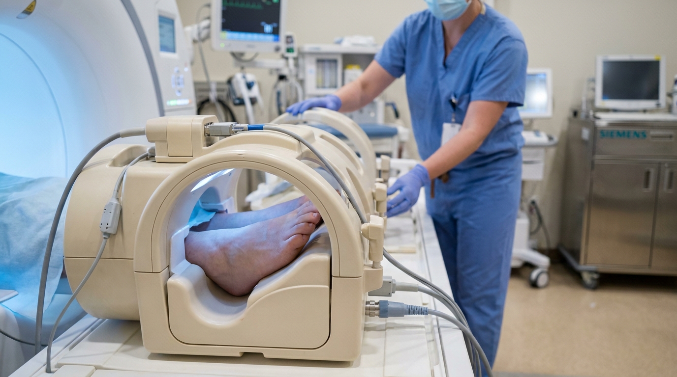
For Doctors in a Hurry
- Researchers investigated whether Pacinian corpuscle counts on forefoot MRI correlate with the severity of large-fiber dysfunction in diabetic sensorimotor polyneuropathy.
- This study evaluated 39 patients with type 2 diabetes and confirmed polyneuropathy using forefoot MRI and nerve conduction studies.
- Patients with severe disease had significantly lower corpuscle counts (54.1) than those with mild-to-moderate impairment (146.1; p < 0.001).
- The researchers concluded that Pacinian corpuscle loss strongly correlates with reduced sural sensory nerve action potential amplitudes (ρ = 0.638).
- MRI-based quantification of these mechanoreceptors may serve as a noninvasive biomarker for assessing large-fiber sensory dysfunction in clinical practice.
Visualizing the Structural Decline of Large-Fiber Sensation
Diabetic sensorimotor polyneuropathy remains a leading cause of functional disability, yet the clinical assessment of sensory recovery and nerve integrity often lacks a definitive diagnostic algorithm [1, 2]. While intraepidermal nerve fiber density via skin biopsy is the established benchmark for small-fiber assessment, evaluating large-fiber involvement typically relies on nerve conduction studies to measure functional loss [3]. These large-diameter fibers terminate in specialized mechanoreceptors known as Pacinian corpuscles, which are essential for detecting high-frequency vibration and maintaining postural stability [4, 5]. Because the structural degradation of these corpuscles may precede or mirror the progression of neuropathic pain and sensory deficits [5], researchers recently investigated whether routine forefoot magnetic resonance imaging can bridge the gap between these structural changes and established electrophysiological measures of nerve dysfunction.
The researchers evaluated 39 patients with type 2 diabetes and neurologically confirmed diabetic sensorimotor polyneuropathy, comprising a cohort with a mean age of 67.9 ± 13.5 years that included 29 males. To investigate the structural correlates of nerve damage, the study utilized forefoot magnetic resonance imaging (MRI) to quantify Pacinian corpuscles (encapsulated nerve endings in the skin responsible for sensitivity to vibration and pressure). These counts were meticulously assessed in both the subcutaneous and deep regions of each digit, providing a localized map of mechanoreceptor density in areas highly susceptible to neuropathic changes. To validate these imaging findings against established clinical standards, participants underwent nerve conduction studies of the tibial, peroneal, and sural nerves. These electrophysiological assessments included the measurement of compound muscle action potentials (the summated electrical response of a muscle to nerve stimulation) and sensory nerve action potentials (the electrical activity propagating along sensory nerves). By comparing these functional metrics with the MRI-derived data, the researchers sought to determine if the physical depletion of Pacinian corpuscles accurately reflects the severity of large-fiber sensory impairment.
Correlation Between Imaging and Electrophysiology
To evaluate the relationship between structural imaging and functional nerve health, the researchers utilized Spearman's rank correlation analysis (a statistical method that measures the strength and direction of association between two ranked variables). This analysis revealed a strong positive association between total Pacinian corpuscle counts and the electrical signal strength of the sural nerve (ρ = 0.638, p < 0.001). This correlation indicates that the depletion of Pacinian corpuscles visualized on MRI serves as a reliable structural indicator of functional decline in large-fiber sensory nerves, providing clinicians with a quantifiable metric that mirrors traditional electrophysiological data. To further delineate the clinical utility of these findings, the study cohort was stratified by disease severity based on motor and sensory amplitudes alongside age-adjusted conduction velocities. This classification identified 10 patients as having mild-to-moderate diabetic sensorimotor polyneuropathy, while 29 patients were classified as having severe disease. In the severe group, the loss of mechanoreceptors was particularly pronounced, closely mirroring the profound electrophysiological deficits observed during nerve conduction testing.
Differentiating Disease Severity Through Structural Markers
The researchers identified a clear divergence in structural and functional markers when comparing patients by disease stage. Among the 29 patients classified with severe diabetic sensorimotor polyneuropathy, 86.1% exhibited abolished sural sensory nerve action potential amplitudes, indicating a profound loss of large-fiber sensory function. This functional collapse was mirrored by a significant reduction in mechanoreceptor density on imaging. The total Pacinian corpuscle counts were significantly lower in severe cases compared to the mild-to-moderate group (54.1 ± 40.6 versus 146.1 ± 43.2; p < 0.001), demonstrating that the physical depletion of these receptors is a hallmark of advanced nerve pathology. Electrophysiological data further quantified this stratification, showing that patients with severe disease had substantially reduced or absent sural sensory nerve action potential amplitudes, with a median of 0.1 µV (range: 0.1 to 1.7 µV). In contrast, the 10 patients with mild-to-moderate disease demonstrated both higher Pacinian corpuscle counts (p < 0.001) and higher sural amplitudes, which showed a median of 2.3 µV (range: 0.9 to 11.4 µV). These findings indicate that MRI-based quantification of Pacinian corpuscles can reliably differentiate between stages of polyneuropathy, providing clinicians with a structural metric that corresponds directly to the degree of electrophysiological impairment.
Clinical Utility as a Noninvasive Biomarker
The integration of forefoot MRI into the diagnostic workup for diabetic sensorimotor polyneuropathy offers a structural perspective that has historically been difficult to quantify without invasive measures. The study demonstrates that forefoot MRI-detected Pacinian corpuscle counts closely correlate with large-fiber sensory function, allowing clinicians to directly visualize the physical state of the peripheral sensory apparatus. This structural assessment mirrors the functional deficits identified in nerve conduction studies, where the loss of these receptors aligns with the reduction or abolition of sural sensory nerve action potential amplitudes. For the practicing physician, these findings suggest that MRI-based Pacinian corpuscle quantification may serve as a noninvasive imaging biomarker for polyneuropathy severity, providing an objective metric to grade disease progression. This approach complements established clinical and electrophysiological methods, offering a reliable alternative when traditional nerve conduction studies are inconclusive, poorly tolerated, or unavailable. Because the researchers found such a distinct separation in corpuscle counts between mild-to-moderate and severe cases, this quantification method holds practical utility for monitoring disease stabilization or decline over time, potentially informing more precise management strategies for patients with diabetic sensory dysfunction.
References
1. John A, Rossettie S, Rafael J, Cox C, Ducic I, MacKay BJ. Clinical Assessment of Pain and Sensory Function in Peripheral Nerve Injury and Recovery: A Systematic Review of Literature. Archives of Plastic Surgery. 2022. doi:10.1055/s-0042-1748658
2. Boulton AJ, Malik RA, Arezzo JC, Sosenko JM. Diabetic Somatic Neuropathies. Diabetes Care. 2004. doi:10.2337/diacare.27.6.1458
3. Baszyńska–Wilk M, Nowacka-Gotowiec M, Moszczyńska E. Diabetic Peripheral Neuropathy in Children and Adolescents - Prevalence, Diagnostic Methods and Risk Factors. Journal of Clinical Research in Pediatric Endocrinology. 2025. doi:10.4274/jcrpe.galenos.2025.2025-3-29
4. Shell CE, Christie B, Marasco PD, Charkhkar H, Triolo RJ. Lower-Limb Amputees Adjust Quiet Stance in Response to Manipulations of Plantar Sensation. Frontiers in Neuroscience. 2021. doi:10.3389/fnins.2021.611926
5. García-Mesa Y, Feito J, Gonzalez-gay M, et al. Involvement of Cutaneous Sensory Corpuscles in Non-Painful and Painful Diabetic Neuropathy. Journal of Clinical Medicine. 2021. doi:10.3390/jcm10194609