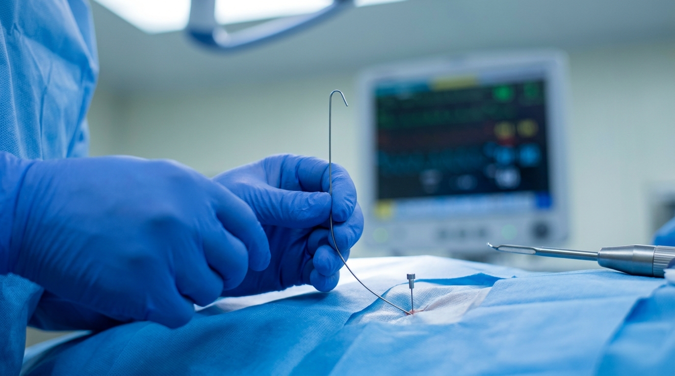
For Doctors in a Hurry
- Surgeons lack consensus on the optimal surgical approach for localizing and resecting small, hilar-proximal, ground-glass opacity dominant lung nodules.
- This multicenter retrospective study evaluated 79 patients with subpleural nodules under 2 centimeters undergoing preoperative CT-guided hookwire localization and wedge resection.
- Localization succeeded in all 79 cases with a median operative time of 15 minutes and no local recurrences after 17.3 months.
- The researchers concluded that this localization strategy effectively facilitates parenchyma-sparing wedge resection for small, centrally located, ground-glass opacity dominant pulmonary nodules.
- This technique allows clinicians to perform lung-preserving surgery for complex hilar-proximal nodules while maintaining low complication rates and short hospital stays.
Parenchyma Preservation for Hilar-Proximal Pulmonary Nodules
The management of early-stage non-small cell lung cancer continues to evolve toward sublobar resection, as evidence suggests wedge resection for nodules 2 cm or smaller can yield oncological outcomes comparable to lobectomy while better preserving pulmonary function [1, 2]. Although video-assisted thoracoscopic surgery (VATS) is standard for these procedures, the intraoperative identification of small, non-palpable lesions remains a significant technical hurdle, especially for those near the pulmonary hilum [3, 4]. These hilar-proximal nodules present unique anatomical challenges that often preclude simple wedge techniques, forcing a choice between more invasive segmentectomy or a parenchyma-sparing approach that risks inadequate margins [5, 6, 7, 8]. A recent multicenter study evaluates a specific localization and dissection strategy designed to facilitate lung-sparing surgery for these difficult-to-reach nodules.
Targeting Small Hilar Lesions
A multicenter retrospective analysis conducted from January 2023 to July 2025 evaluated a surgical strategy for 79 patients with challenging subpleural pulmonary nodules located near the hilum. These hilar-proximal lesions are clinically problematic because their deep anatomical position often necessitates a more extensive resection to ensure adequate margins, even for small, radiographically indolent nodules. To isolate a specific patient population, the study applied strict inclusion criteria based on preoperative imaging. All included nodules measured 2 cm or less in diameter, with the cohort exhibiting a median nodule size of 7 mm (range 6 to 20 mm). Furthermore, eligibility required a consolidation-to-tumor ratio (CTR) of 0.25 or less. The CTR, which quantifies the solid component of a nodule on computed tomography (CT), is a key radiographic biomarker; a low CTR indicates a ground-glass opacity-dominant lesion, a feature often associated with early-stage adenocarcinoma where a parenchyma-sparing wedge resection is considered oncologically appropriate if the lesion can be accurately localized and completely excised.
Localization and Surgical Technique
Precise preoperative mapping was foundational to the surgical intervention, with clinicians achieving a 100 percent success rate in localizing all 79 nodules using CT-guided hookwire placement. To access these deep-seated lesions, the team employed either a transfissural approach, which passes the wire through the natural fissure between lung lobes, or a traversing-lobe approach, where the wire passes through one lobe to mark a nodule in an adjacent one. This level of precision is essential for hilar-proximal nodules, whose proximity to major vessels and central airways makes them nearly impossible to identify by manual palpation or direct visualization during surgery. Following localization, patients underwent uniportal VATS wedge resection. To maximize the preservation of healthy lung tissue, the surgical team primarily used ultrasonic dissection, a technique that employs high-frequency vibrations to cut and coagulate tissue with minimal lateral thermal damage. This method facilitated meticulous dissection around the nodule with limited use of surgical staplers, which can crush and remove a larger margin of healthy parenchyma. This approach allowed surgeons to navigate complex hilar anatomy and excise the target nodules while retaining the maximum possible volume of functional lung tissue.
Procedural Efficiency and Safety Profile
The combination of precise localization and targeted dissection resulted in high procedural efficiency. The median operative time was 15.0 minutes (range 10 to 28 minutes), a duration enabled by the clear roadmap provided by the hookwire. The study reported that no other major complications occurred intraoperatively, indicating the strategy is a viable option for these anatomically complex resections. The localization procedure itself was associated with a favorable safety profile, characterized by minor, manageable events. Following hookwire placement, minor asymptomatic pneumothorax occurred in 12 patients and small pulmonary hemorrhage in 10 patients, neither of which required intervention before surgery. Postoperatively, the most common complication was a persistent air leak lasting more than 72 hours, observed in 10 patients, an outcome not unexpected given the dissection of parenchyma near the hilum. Overall recovery was swift, with a median postoperative hospital stay of 3 days (range 2 to 6 days), consistent with established minimally invasive thoracic procedures.
Pathological Outcomes and Long-Term Control
Histopathological analysis of the 79 resected nodules confirmed the malignant or pre-malignant nature of most lesions, validating the decision to intervene. The findings included 9 cases of adenocarcinoma in situ, 53 cases of minimally invasive adenocarcinoma, and 14 cases of invasive adenocarcinoma, alongside 3 benign lesions. The high proportion of early-stage malignancies (adenocarcinoma in situ and minimally invasive adenocarcinoma) underscores the clinical rationale for a parenchyma-sparing approach, as these lesions are well-suited for curative resection with less extensive surgery. The oncological efficacy of this technique was supported by follow-up data. Over a median follow-up of 17.3 months, no local recurrences or deaths were detected. These outcomes suggest that for carefully selected small, ground-glass opacity-dominant, hilar-proximal nodules, this combined localization and dissection strategy can achieve complete oncological resection while maximizing lung preservation.
References
1. Hu J, Chen J, Chen C, Zhong W, Geng Q. [Wedge Resection of Pulmonary Nodules (≤2 cm):
A Consensus Statement by Specialists of Thoracic Surgery (2023 Edition)].. Zhongguo fei ai za zhi = Chinese journal of lung cancer. 2023. doi:10.3779/j.issn.1009-3419.2023.102.17
2. Cardillo G, Petersen RH, Ricciardi S, et al. European guidelines for the surgical management of pure ground-glass opacities and part-solid nodules: Task Force of the European Association of Cardio-Thoracic Surgery and the European Society of Thoracic Surgeons. European Journal of Cardio-Thoracic Surgery. 2023. doi:10.1093/ejcts/ezad222
3. Sedrakyan A, Meulen JVD, Lewsey J, Treasure T. Video assisted thoracic surgery for treatment of pneumothorax and lung resections: systematic review of randomised clinical trials. BMJ. 2004. doi:10.1136/bmj.38243.440486.55
4. Neijenhuis LKA, Muynck LDAND, Bijlstra OD, et al. Near-Infrared Fluorescence Tumor-Targeted Imaging in Lung Cancer: A Systematic Review. Life. 2022. doi:10.3390/life12030446
5. Tang X, Jian H, Guan Y, Miao J, Liang X. Computed tomography-guided localization for multiple pulmonary nodules: a meta-analysis.. Videosurgery and Other Miniinvasive Techniques. 2021. doi:10.5114/wiitm.2021.104199
6. Chen K, Niu Z, Jin R, et al. Three-dimensional reconstruction computed tomography in thoracoscopic segmentectomy: a randomized controlled trial.. European journal of cardio-thoracic surgery : official journal of the European Association for Cardio-thoracic Surgery. 2024. doi:10.1093/ejcts/ezae250
7. Sun Z, Cheng H, Su J, Sun Q. Preoperative localization for pulmonary nodules: a meta-analysis of coil and liquid materials.. Minimally invasive therapy & allied technologies : MITAT : official journal of the Society for Minimally Invasive Therapy. 2024. doi:10.1080/13645706.2024.2337073
8. Bade BC, Blasberg JD, Mase VJ, et al. A guide for managing patients with stage I NSCLC: deciding between lobectomy, segmentectomy, wedge, SBRT and ablation—part 3: systematic review of evidence regarding surgery in compromised patients or specific tumors. Journal of Thoracic Disease. 2022. doi:10.21037/jtd-21-1825