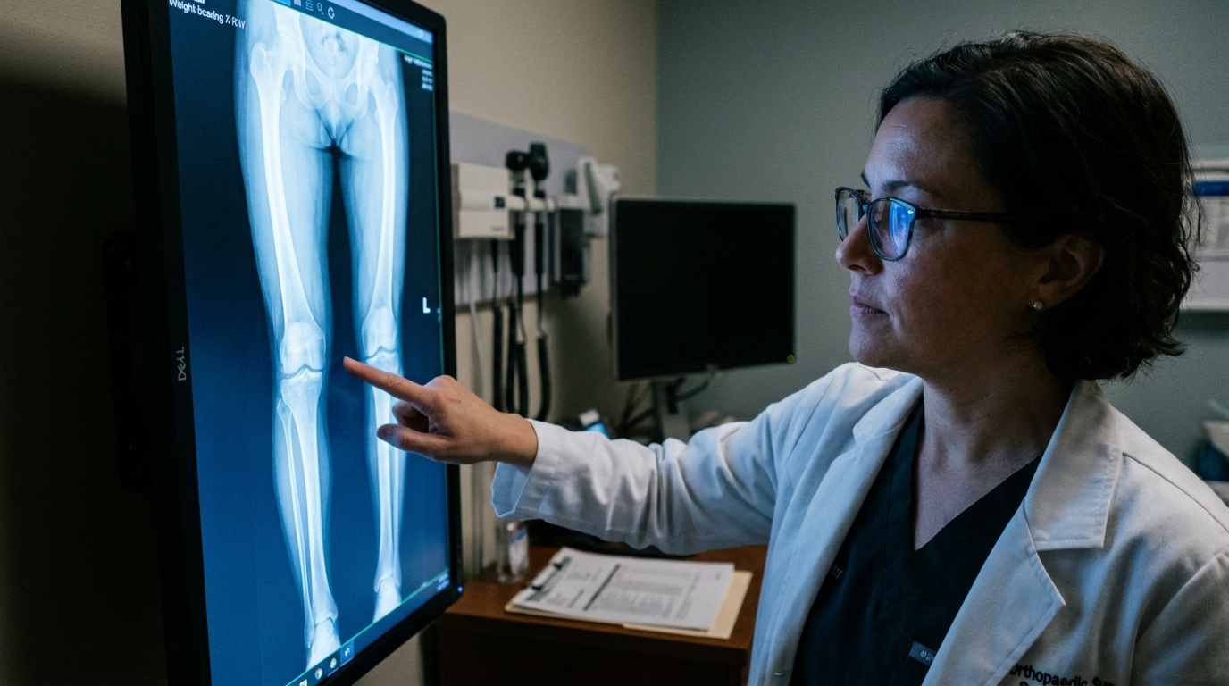
For Doctors in a Hurry
- Clinicians need to determine if long bone bowing influences the classification of knee alignment phenotypes during surgical planning.
- The researchers retrospectively analyzed 192 knees to evaluate the relationship between bone bowing and standard radiographic alignment parameters.
- Tibial bowing correlated with alignment angles, with valgus knees showing a significantly higher mean bowing of 2.64 degrees.
- The study concludes that bone bowing significantly alters knee classification, often masking a more neutral underlying joint alignment.
- Surgeons should account for tibial and femoral bowing to avoid misclassifying knee phenotypes when planning joint resurfacing procedures.
Coronal plane alignment serves as a primary determinant of knee biomechanics, influencing joint loading, ligament tension, and the long-term risk of secondary joint degeneration [1]. In patients with genu valgum (knock-kneed deformity), increased stress on the anteromedial bundle of the anterior cruciate ligament can compromise joint stability and elevate the risk of non-contact injuries [2]. While individual knee phenotyping (the categorization of patients based on their unique anatomical traits) has recently transformed alignment paradigms in arthroplasty, the influence of extra-articular factors like long-bone morphology remains a significant challenge for surgical planning [3, 1]. Precise assessment of these deformities is critical, as minor initial malalignments may lead to unpredictable outcomes or rebound phenomena following guided growth interventions [4]. A recent retrospective analysis of 192 knees now investigates how metaphyseal bowing specifically contributes to the natural history of valgus knee phenotypes.
The researchers conducted a retrospective analysis of 192 knees between April 2024 and September 2025 to determine how long bone morphology influences established classification systems. Central to this investigation was the Coronal Plane Alignment of the Knee (CPAK), a framework that characterizes knee phenotypes by combining the arithmetic hip-knee-ankle angle (aHKA), which measures overall limb alignment from the center of the hip to the center of the ankle, and joint line obliquity (JLO), which describes the tilt of the joint surface relative to the floor. To isolate the impact of bone shape, the study measured tibial bowing (tBOW) and femoral bowing (fBOW) angles defined by the relationship between the proximal and distal metadiaphyseal axes (the longitudinal centerlines of the bone shafts just before they expand into the joint complex). This distinction is vital because it separates the curvature of the bone shaft from the geometry of the joint itself.
The study utilized several key radiographic parameters to quantify alignment, including the lateral distal femoral angle (LDFA) and the medial proximal tibial angle (MPTA). To understand the true contribution of bone curvature, the authors calculated a corrected MPTA (the MPTA without the influence of tBOW) and a corrected LDFA (the LDFA without the influence of fBOW). The data revealed that tibial bowing significantly correlated with both MPTA (r = 0.56, P < 0.001) and aHKA (r = 0.47, P < 0.001), suggesting that what often appears as joint-level malalignment is frequently a function of the tibial shaft's curvature. Similarly, femoral bowing showed strong correlations with LDFA (r = -0.60, P < 0.001) and JLO (r = -0.50, P < 0.001), indicating that the femoral metadiaphyseal shape is a primary driver of distal femoral geometry and joint line orientation.
Bowing as a Driver of Valgus Phenotypes
The researchers identified distinct morphological patterns across different alignment groups, noting that the degree of tibial curvature varies significantly depending on the overall limb phenotype. Specifically, valgus knees exhibited a mean tibial bowing of 2.64 degrees, which was substantially higher than the curvature observed in other cohorts. In contrast, neutral knees demonstrated a mean tibial bowing of 0.71 degrees, while varus knees showed a mean tibial bowing of -0.14 degrees. The difference in mean tibial bowing between the valgus, neutral, and varus groups was statistically significant (P < 0.001), suggesting that the presence of a valgus deformity is frequently associated with a specific diaphyseal shape rather than being isolated to the joint surface itself. This finding implies that clinicians should look beyond the joint capsule when assessing the etiology of valgus malalignment.
The study further clarified how these bony curvatures influence the overall geometry of the lower limb. The researchers found that medial tibial bowing and lateral femoral bowing increase the valgus arithmetic hip-knee-ankle angle (aHKA), which is a measure of the overall limb alignment calculated from radiographic landmarks. Furthermore, these bowing patterns contribute to an apex-proximal joint line obliquity (JLO), a term describing a joint line that tilts upward toward the lateral aspect of the knee. For the clinician, this indicates that a significant portion of what is traditionally classified as a valgus knee phenotype may actually be driven by extra-articular factors. Recognizing that the normal apex-distal target joint line (a joint line that tilts downward toward the lateral side) is displaced in part by the degree of the bow is critical for surgeons, as it suggests that standard intra-articular corrections may not fully address the underlying structural deformity.
Phenotypic Reclassification and Surgical Implications
The clinical impact of extra-articular curvature becomes most evident when researchers apply an arithmetic correction to account for the influence of long bone bowing on standard alignment metrics. By mathematically removing the effect of tibial bowing from the calculations, the researchers found that 71.3% of all valgus knees were reclassified toward more neutral alignment classifications. This shift suggests that for a vast majority of patients, the perceived valgus deformity is not a product of the joint surface itself but is instead driven by the underlying diaphyseal shape. This reclassification was particularly pronounced across specific Coronal Plane Alignment of the Knee (CPAK) phenotypes. Specifically, arithmetic correction for tibial bowing reclassified 60.3% of CPAK type 3 cases, 100% of CPAK type 6 cases, and 100% of CPAK type 9 cases, moving them into more neutral categories.
These findings indicate that tibial and femoral bowing fundamentally alter CPAK classification, and accounting for these curvatures returns many valgus knees to a neutral, apex-distal alignment. For the practicing surgeon, this distinction is vital because the presence of bowing means the normal apex-distal target joint line is displaced in part by the amount of the bow. While small degrees of bowing do not necessarily necessitate an extra-articular osteotomy (a surgical procedure to realign the bone by cutting and repositioning it), they must be carefully considered by surgeons performing knee resurfacing who are guided by collateral ligament positions. Failure to recognize that the deformity is extra-articular may lead to inappropriate intra-articular resections or ligamentous releases, as the surgeon must distinguish between a true joint-level malalignment and the displacement caused by the metadiaphyseal bow. This study underscores the need for a more holistic radiographic assessment that accounts for the entire length of the femur and tibia before committing to a surgical plan.
References
1. Sadoghi P, Koutp A, Hirschmann MT. Unlocking the scientific and clinical value of knee phenotyping beyond knee arthroplasty. Knee Surgery Sports Traumatology Arthroscopy. 2025. doi:10.1002/ksa.70142
2. Jian X, Sun D, Xu Y, et al. Running-Induced Fatigue Exacerbates Anteromedial ACL Bundle Stress in Females with Genu Valgum: A Biomechanical Comparison with Healthy Controls. Sensors. 2025. doi:10.3390/s25154814
3. Pratobevera A, Seil R, Ménétrey J. Joint line and knee osteotomy. EFORT Open Reviews. 2024. doi:10.1530/eor-24-0037
4. Stief F, Holder J, Braun S, et al. Relevance of instrumented gait analysis in the prediction of the rebound phenomenon after guided growth intervention. Scientific Reports. 2024. doi:10.1038/s41598-024-66169-9