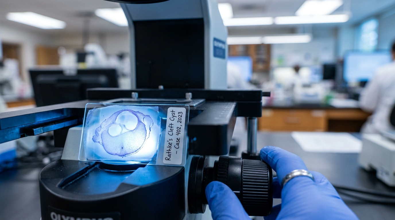
For Doctors in a Hurry
- Researchers investigated whether specific molecular markers identified in mice could explain the clinical symptoms and pathogenesis of human Rathke's cleft cysts.
- This retrospective analysis evaluated 108 histopathologically confirmed cases of Rathke's cleft cysts resected at three medical centers between 2011 and 2023.
- FOXJ1 positivity significantly correlated with hypopituitarism (p = 0.040), while absent TUBA1A expression was associated with arginine vasopressin deficiency (p = 0.042).
- The study confirms these six markers are expressed in humans, suggesting KRT8 and FOXA1 may help distinguish these cysts from craniopharyngiomas.
- These molecular profiles may improve diagnostic precision and provide prognostic insights into endocrine dysfunction for patients with symptomatic sellar lesions.
Molecular drivers of endocrine dysfunction in Rathke’s cleft cysts
Rathke’s cleft cysts are benign sellar lesions often discovered incidentally during imaging for unrelated symptoms, yet they carry a significant risk of morbidity when they compress the pituitary gland or hypothalamus [1]. While many remain asymptomatic, symptomatic cases frequently present with headaches, visual disturbances, and endocrine dysfunction, including a 14% prevalence of permanent arginine vasopressin deficiency (the underlying cause of central diabetes insipidus, characterized by an inability to concentrate urine) following surgical intervention [2]. Distinguishing these cysts from more aggressive lesions like craniopharyngiomas remains a diagnostic challenge, as both share similar anatomical locations and clinical presentations [3]. Current management relies heavily on clinical symptoms and magnetic resonance imaging, but the underlying molecular mechanisms driving cyst pathogenesis and associated hormonal deficits are poorly understood [4]. To address this gap, researchers recently conducted a retrospective analysis evaluating how specific molecular markers correlate with these clinical manifestations, aiming to improve diagnostic precision and better predict postoperative endocrine outcomes.
Translating murine markers to human pathology
To understand the biological drivers of Rathke’s cleft cysts, investigators turned to a set of proteins previously linked to pituitary development. Building on prior research in Isl-1 knockout mice (a laboratory model genetically engineered to lack a key regulatory protein, allowing scientists to study pituitary gland formation), researchers identified six molecular markers potentially involved in cyst pathogenesis: KRT8, TUBA1A, SOX2, SOX9, FOXA1, and FOXJ1. The current study sought to determine whether these markers are present in human tissue and if their expression correlates with specific clinical manifestations, such as hypopituitarism or arginine vasopressin deficiency. To investigate these associations, the researchers conducted a retrospective analysis of 108 histopathologically confirmed cases resected between 2011 and 2023 across three medical centers. The team utilized immunofluorescence staining, a technique that uses fluorescently labeled antibodies to visualize specific proteins within tissue samples, to evaluate the expression of the six identified markers. This multicenter cohort allowed for a robust comparison of molecular profiles against a variety of clinical and diagnostic variables, including magnetic resonance imaging findings and epithelial morphology (the specific cellular arrangement of the cyst lining). Using chi-square and Fisher’s exact tests, the researchers mapped these molecular signatures to the physical characteristics of the cysts and the patients' preoperative presentations. By doing so, the study aimed to identify objective biological indicators that might predict the clinical course of the disease or assist in the differential diagnosis of sellar masses.
Expression prevalence and diagnostic differentiation
Tissue analysis confirmed that the molecular markers identified in animal models are highly prevalent in human Rathke’s cleft cysts. KRT8 (Keratin 8) was expressed in 100% of the 108 samples, establishing it as a universal marker within this cohort. The researchers also observed a high frequency of TUBA1A (Tubulin Alpha 1a) expression, which was present in 90.7% of the human samples. These high expression rates suggest that these structural proteins are fundamental components of the cyst's cellular architecture. The remaining four markers demonstrated substantial, though more variable, expression across the patient cohort. The researchers reported that SOX2 was expressed in 75.9% of samples, while SOX9 expression was identified in 76.9% of the cases. Furthermore, FOXJ1 (Forkhead Box J1) showed an expression rate of 84.3%, and FOXA1 (Forkhead Box A1) was present in 55.6% of the human samples. These variations in protein expression provide a molecular map that may eventually help clinicians categorize these lesions based on their specific biological signatures rather than relying solely on gross morphology. Beyond characterizing the cysts themselves, the findings offer a practical tool for resolving diagnostic ambiguity in the sellar region. Distinguishing benign cysts from craniopharyngiomas (more aggressive epithelial tumors that often require wider surgical resection and closer postoperative surveillance) can be challenging when using magnetic resonance imaging alone. The researchers found that KRT8 and FOXA1 staining may aid in distinguishing Rathke’s cleft cysts from craniopharyngiomas. By integrating these molecular markers into routine pathological evaluations, surgical teams could achieve greater diagnostic certainty, directly informing the extent of surgical intervention and long-term management strategies.
Correlating molecular profiles with clinical morbidity
The most clinically actionable insights emerged when researchers correlated protein expression profiles with specific endocrine deficits. Symptomatic Rathke’s cleft cysts frequently cause hypopituitarism and arginine vasopressin deficiency, the latter characterized by inadequate antidiuretic hormone secretion leading to profound polyuria and polydipsia. In this cohort, the analysis revealed that FOXJ1 positivity was significantly associated with hypopituitarism (p = 0.040), suggesting that the presence of this transcription factor may serve as a biological indicator of anterior pituitary dysfunction. Conversely, the absence of TUBA1A expression was significantly associated with arginine vasopressin deficiency (p = 0.042). For practicing endocrinologists and neurosurgeons, these findings provide specific biological insights into why histologically similar cysts can produce vastly different hormonal presentations, raising the possibility that future tissue biopsies could help predict a patient's risk for permanent endocrine failure. The study also identified a strong link between molecular markers and the physical structure of the cyst wall, noting that SOX9 expression was significantly associated with single-layered epithelial morphology (p = 0.001). However, these molecular signatures did not extend to all clinical parameters. There were no significant associations found between marker expression and imaging findings on magnetic resonance imaging, nor were there any significant associations between marker expression and visual disturbances or headache. This lack of correlation suggests a clear mechanistic divide: while molecular markers like FOXJ1 and TUBA1A are intimately tied to specific endocrine disruptions, neurological symptoms such as vision loss or cephalalgia are likely driven purely by the physical mass effect of the cyst compressing adjacent structures.
References
1. Hwang Y, Ku C. Revisiting Pituitary Incidentalomas: Insights from Prevalence Data and Consensus Recommendations. Endocrinology and Metabolism. 2025. doi:10.3803/EnM.2025.2688
2. Fountas A, Coulden A, Fernández-García S, Tsermoulas G, Allotey J, Karavitaki N. Central diabetes insipidus (vasopressin deficiency) after surgery for pituitary tumours: a systematic review and meta-analysis. European Journal of Endocrinology. 2024. doi:10.1093/ejendo/lvae084
3. Alboqami MN, Albaiahy A, Bukhari BH, et al. Craniopharyngioma: A comprehensive review of the clinical presentation, radiological findings, management, and future Perspective. Heliyon. 2024. doi:10.1016/j.heliyon.2024.e32112
4. Brinkmeier ML, Bando H, Camarano AC, et al. Rathke’s cleft-like cysts arise from Isl1 deletion in murine pituitary progenitors. Journal of Clinical Investigation. 2020. doi:10.1172/jci136745