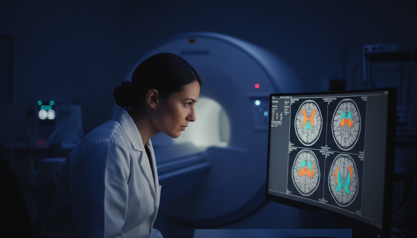
For Doctors in a Hurry
- Researchers investigated structural and metabolic brain changes across three phases of epilepsy development following prolonged seizures, known as status epilepticus.
- The study analyzed forty-eight rats using MRI to measure dorsal hippocampal volume and N-acetylaspartate, a marker of neuronal health, over thirty days.
- Seizure-induced rats showed reduced hippocampal volumes and N-acetylaspartate levels at all post-baseline scans compared to controls (p < 0.001).
- Acute hippocampal damage occurs within forty-eight hours of status epilepticus, followed by a pattern of partial but incomplete structural recovery.
- Tracking these noninvasive markers could help establish surrogate endpoints for future clinical trials aiming to prevent focal epilepsy.
Tracking the Hidden Phases of Epileptogenesis
Temporal lobe epilepsy often develops after an initial severe brain insult, such as status epilepticus, which triggers a cascade of neurological changes known as epileptogenesis [1, 2]. This process typically unfolds in three distinct phases: an acute injury, a clinically silent latent period, and a chronic phase characterized by spontaneous recurrent seizures [3]. During this progression, patients frequently develop significant cognitive and psychiatric comorbidities that severely complicate clinical management [4]. Because the structural and metabolic alterations driving this transformation occur long before the first spontaneous seizure, identifying non-invasive biomarkers to track these hidden changes remains a major clinical challenge [2]. A recent study evaluates how these structural and metabolic markers evolve over time, providing data that could eventually help clinicians monitor disease progression and time targeted interventions before chronic epilepsy takes hold.
Mapping the Timeline of Hippocampal Damage
To evaluate longitudinal structural and metabolic changes across the three phases of epileptogenesis, researchers analyzed 48 male eight-week-old Wistar rats. The animals were assigned to either a sham-control group or an experimental group subjected to the pilocarpine model of temporal lobe epilepsy, a standard experimental method used to induce status epilepticus and subsequent chronic seizures. To capture the evolving pathology, the investigators acquired T2-weighted images and proton magnetic resonance spectra, a non-invasive imaging technique that measures brain metabolites. All imaging was performed using a 3 T MRI clinical scanner (Philips Achieva) equipped with an animal coil, allowing the team to utilize clinical-grade technology to track disease progression. The study specifically measured dorsal hippocampal volumes to assess structural atrophy and calculated the ratio of total N-acetylaspartate to total creatine (tNAA/tCr), a well-established marker of neuronal integrity and metabolic health. These measurements were taken at four distinct time points to map the disease trajectory. The initial scans occurred at baseline, before any treatments were administered. Subsequent measurements were taken at 48 hours to represent the acute phase of injury, followed by scans at 15 days to capture the clinically silent period. Finally, the team evaluated the animals at 30 days, marking the beginning of the chronic phase.
To evaluate the longitudinal data, the researchers built generalized linear mixed effects models, a statistical approach used to analyze repeated measurements from the same subjects over time while accounting for individual variability. These models tested differences in dorsal hippocampal volume and tNAA/tCr ratios, including group assignment and MRI scan timing as main effects. Prior to the experimental intervention, the pilocarpine-induced and control animals showed similar baseline dorsal hippocampal volumes and tNAA/tCr ratios (both p > 0.1), establishing that both cohorts began with equivalent structural and metabolic profiles. Following the induction of seizures, the experimental group experienced immediate neurological deterioration. The imaging revealed that acute dorsal hippocampal volume loss and hippocampal neuronal dysfunction were present as early as 48 hours post-status epilepticus. This structural and metabolic damage persisted throughout the entire observation period. Specifically, the experimental animals showed reduced dorsal hippocampal volumes at all post-baseline MRI scans (all p < 0.001) when compared to controls. Furthermore, metabolic integrity remained compromised, as the experimental group demonstrated reduced tNAA/tCr ratios at all post-baseline scans (all p < 0.001) compared to controls. In stark contrast, normal physiological development proceeded without interruption in the unexposed cohort, as there were no changes over time in dorsal hippocampal volume or tNAA/tCr ratios in the sham-controls (all p > 0.4).
Partial Recovery During the Silent and Chronic Phases
While the initial insult caused immediate structural and metabolic declines, the researchers observed a dynamic shift as the disease progressed. An intragroup analysis tracking the affected animals over time revealed a partial rebound in brain structure, showing that dorsal hippocampal volumes significantly increased at 15 and 30 days (all p < 0.001) when compared to the 48-hour mark. Similarly, metabolic function showed signs of a rebound, with tNAA/tCr ratios significantly increasing at 15 and 30 days (all p < 0.001) compared to the acute phase. This trajectory indicates that the acute damage is followed by a pattern of gradual recovery throughout the silent and chronic phases of epileptogenesis. However, this rebound was ultimately incomplete. Despite the increases at 15 and 30 days, both the dorsal hippocampal volumes and tNAA/tCr ratios in the experimental group remained lower than their baseline scans, indicating a permanent structural and metabolic deficit. For clinicians managing patients after severe neurological insults, these findings highlight a critical window of ongoing biological changes before the onset of spontaneous seizures. Understanding the course of these non-invasive markers of hippocampal sclerosis may help establish surrogate endpoints for future clinical trials. By utilizing standard clinical MRI technology to track hippocampal volume and metabolic ratios, physicians could eventually measure whether neuroprotective therapies successfully alter this trajectory during the latent phase, rather than waiting years to observe clinical seizure frequency.
References
1. Wang Y, Wei P, Feng Y, Luo Y, Zhao G. Animal Models of Epilepsy: A Phenotype-oriented Review. Aging and Disease. 2022. doi:10.14336/ad.2021.0723
2. Antmen FM, Matpan E, Dayanc ED, et al. The Metabolic Profile of Plasma During Epileptogenesis in a Rat Model of Lithium–Pilocarpine-Induced Temporal Lobe Epilepsy. Molecular Neurobiology. 2025. doi:10.1007/s12035-025-04719-6
3. Roginskaya AI, Kovalenko AA, Zubareva OE. Effect of pioglitazone on the behavior and expression of genes involved in the regulation of epileptogenesis in a lithium-pylocarpine model of temporal lobe epilepsy in rats. 2023. doi:10.17816/gc623301
4. Mazarati A, Siddarth P, Baldwin RA, Shin D, Caplan R, Sankar R. Depression after status epilepticus: behavioural and biochemical deficits and effects of fluoxetine. Brain. 2008. doi:10.1093/brain/awn117