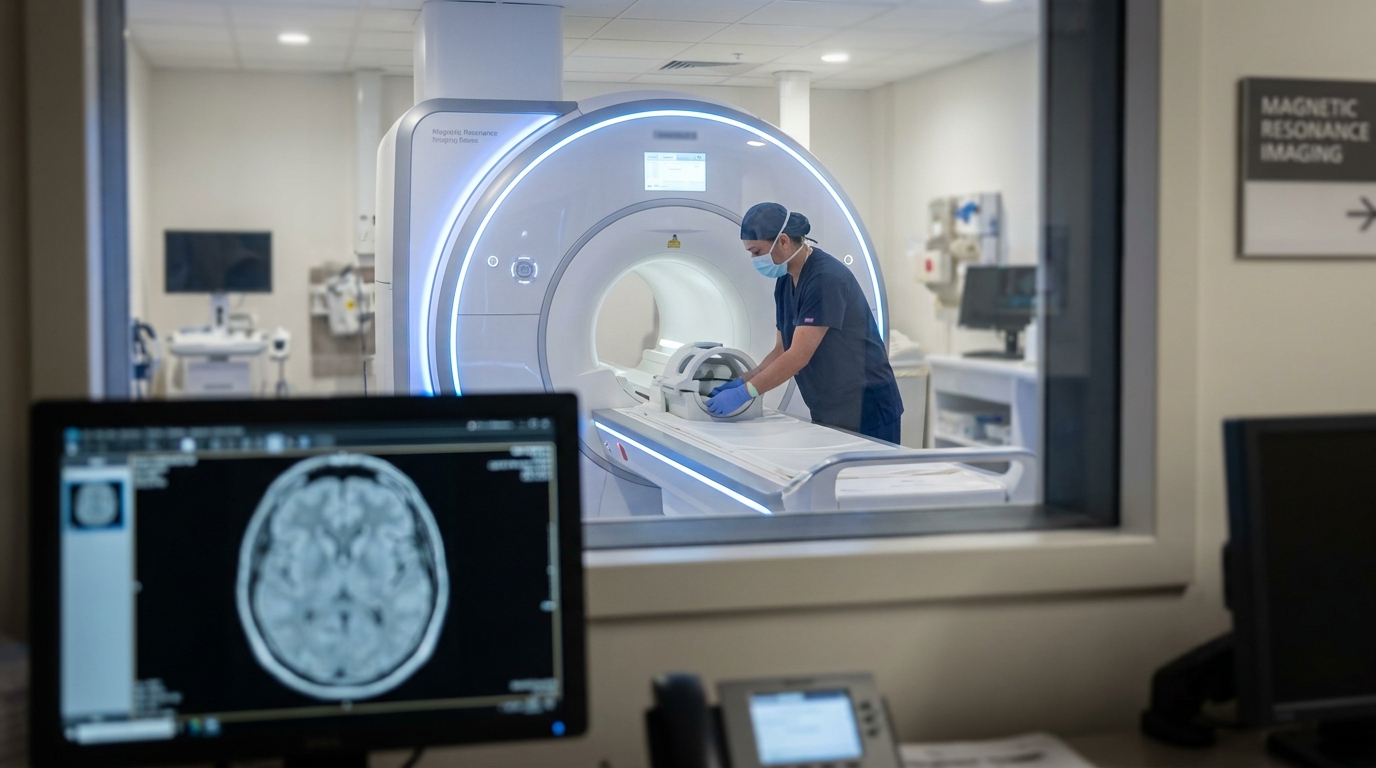
For Doctors in a Hurry
- Traditional group-based analyses often obscure individual neuroanatomical differences in traumatic brain injury because they average across diverse injury patterns.
- Researchers analyzed MRI data from 1,384 patients using normative modeling, which maps individual brain deviations from a healthy baseline.
- Regional convergence of brain deviations did not exceed 34 percent, while median deviations increased from 9 to 22 with severity.
- The study concludes that normative modeling identifies patient-specific cortical and subcortical abnormalities that conventional group-level comparisons frequently overlook.
- Creating individualized morphologic fingerprints may improve prognostic accuracy and help clinicians develop personalized treatment strategies for brain injury.
The Challenge of Neuroanatomical Heterogeneity in Traumatic Brain Injury
Traumatic brain injury remains a leading cause of long-term disability worldwide, contributing significantly to the global burden of neurological morbidity [1]. While clinical management often relies on standardized assessments like the Glasgow Coma Scale, these tools frequently fail to capture the full spectrum of functional and structural anomalies that can persist years after the initial insult [2, 3]. Current neuroimaging protocols struggle with the inherent heterogeneity of injury patterns, making it difficult to establish reliable biomarkers for individual prognosis [4]. This diagnostic gap is particularly evident when group-level averages obscure the unique pathological signatures of single patients. To address this challenge, researchers recently applied normative modeling, a statistical framework that establishes a baseline of typical brain development to flag individual deviations, mapping how each patient's neuroanatomy differs from healthy expectations.
Comparing Group-Level Averages with Individual Deviations
To investigate the structural impact of head trauma, researchers analyzed neuroimaging data from the Enhancing NeuroImaging Genetics through Meta-Analysis (ENIGMA) Consortium Adult Moderate-to-Severe TBI working group. This large-scale analysis included 1,344 participants with traumatic brain injury and 1,111 healthy controls. The study utilized two distinct analytical paths to evaluate brain structure: a traditional case-control approach and normative modeling. By applying both methods to cortical and subcortical magnetic resonance imaging data, the authors sought to determine if individual-level analysis could capture pathological changes typically lost when data are averaged across a large group. The researchers focused on two primary outcome measures: cortical thickness, derived from the Destrieux atlas, and subcortical volumes, calculated using the Freesurfer subcortical atlas.
For the traditional case-control portion of the study, the team employed linear models controlling for age, sex, imaging site, and total intracranial volume. While this standard method identifies areas where the patient group as a whole differs from the control group, it often fails to account for the high degree of anatomical variability seen in clinical practice. To address this limitation, the study performed normative modeling via the Predictive Clinical Neuroscience Portal. This process utilized Bayesian linear regression with likelihood warping, a mathematical technique that maps the expected distribution of brain metrics across a healthy population while adjusting for complex, non-linear data patterns. By using this model, the researchers calculated a Z-score for every brain region in each individual patient, representing exactly how far that specific person's brain volume or thickness deviated from the predicted healthy range. For the practicing physician, this approach shifts the focus from a generalized group average to the unique structural signature of a single patient's injury, potentially guiding more targeted rehabilitation strategies.
The Limitations of Conventional Case-Control Imaging
The clinical management of traumatic brain injury is inherently difficult because the condition is highly heterogeneous, complicating the development of standardized diagnostic and therapeutic approaches. In traditional neuroimaging research, investigators typically rely on case-control designs to identify structural changes. However, these conventional group-based analyses often obscure individual differences by averaging across diverse injury patterns, effectively washing out the unique pathological signatures that a clinician sees in an individual patient. By focusing on the mean difference between a patient cohort and a control group, researchers may overlook the specific structural deviations that drive a particular patient's functional deficits.
In this study, traditional case-control comparisons identified significant group-level differences in 10 cortical regions and 5 subcortical regions. When the data were aggregated, the researchers found consistent structural alterations in several key deep brain structures, specifically the bilateral hippocampus, bilateral thalamus, and right amygdala. These areas are frequently associated with the cognitive and emotional processing deficits observed post-injury. The analysis further localized group-level differences to several regions of the frontal and parietal lobes. Significant findings were reported in the bilateral superior frontal sulcus, bilateral middle frontal sulcus, and bilateral precentral gyrus, as well as the bilateral intraparietal sulcus and bilateral transverse occipital sulcus.
While these results provide a generalized map of where traumatic brain injury impacts the brain, they highlight the limitations of group averages in clinical practice. Because these averages mask the true diversity of injury patterns, a patient could have severe focal damage in a region not listed as significant in a group study. Conversely, they might lack typical hippocampal atrophy despite having a severe injury. This discrepancy underscores why group-level data often fail to translate into precise prognostic tools for individual bedside care.
Mapping the Individualized Morphologic Fingerprint
By comparing individual brain scans to the distribution observed in healthy controls, the researchers utilized normative modeling to capture patient-specific deviations from expected neuroanatomical norms. This analysis revealed a striking lack of regional convergence among patients, demonstrating that traumatic brain injury is a highly idiosyncratic process. Specifically, the data showed that no more than 14% of participants with traumatic brain injury shared an extreme deviation (defined as a Z-score less than -2) in the exact same brain region. This finding suggests that the structural damage clinicians observe on a magnetic resonance imaging scan is rarely localized to the same anatomical coordinates across different patients, even when they share similar injury mechanisms.
The diversity of these structural changes was further evidenced by the fact that every brain region experienced at least one extreme positive or negative deviation across the study population. This indicates that no area of the brain is universally spared or uniquely vulnerable across all cases of head trauma. Because normative modeling detects participant-specific cortical and subcortical abnormalities that conventional group comparisons overlook, it provides a more accurate representation of the true diversity of injury-related morphologic changes. By identifying these unique patterns, the researchers have laid the groundwork for generating a morphologic fingerprint for each patient. For the practicing physician, this means that the absence of atrophy in a common area like the hippocampus does not rule out significant structural pathology elsewhere, as injury signatures are almost entirely individualized.
Injury Severity and Clinical Prognosis
The researchers further examined how individual neuroanatomical deviations correlate with clinical measures of injury severity by stratifying the patient cohort according to Glasgow Coma Scale (GCS) scores. This analysis included three distinct subgroups: patients with mild traumatic brain injury (GCS 13 to 15), those with moderate injury (GCS 9 to 12), and those with severe injury (GCS 3 to 8). The data demonstrated that the median number of structural deviations increased in direct proportion to injury severity. Specifically, patients in the GCS 13 to 15 group exhibited a median of 9 deviations, while those in the GCS 9 to 12 subgroup showed a median of 19 deviations. The most severe cases, represented by the GCS 3 to 8 subgroup, reached a median of 22 deviations. These findings indicate that as the clinical severity of the injury increases, the brain undergoes more widespread structural changes, yet these changes remain highly idiosyncratic to the individual patient.
Despite the increase in the total number of deviations among more severely injured patients, the specific anatomical locations of these abnormalities remained remarkably inconsistent. Even when stratifying by injury severity, regional convergence did not exceed 34% in any subgroup, meaning that no single brain region was affected in more than approximately one-third of patients within the same severity category. This lack of commonality underscores how group averages mask highly individualized morphologic abnormalities as injury severity increases. For the clinician, this suggests that traditional imaging summaries may fail to capture the full extent of pathology in the most vulnerable patients. Generating individualized morphologic fingerprints, a detailed map of patient-specific structural brain changes, may ultimately advance prognostic accuracy and lay the foundation for personalized interventions in clinical practice, allowing for rehabilitation strategies tailored to the specific neuroanatomical damage of each patient.
References
1. Kassebaum NJ, Arora M, Barber RM, et al. Global, regional, and national disability-adjusted life-years (DALYs) for 315 diseases and injuries and healthy life expectancy (HALE), 1990–2015: a systematic analysis for the Global Burden of Disease Study 2015. The Lancet. 2016. doi:10.1016/s0140-6736(16)31460-x
2. Amico F, Koberda JL. Quantitative Electroencephalography Objectivity and Reliability in the Diagnosis and Management of Traumatic Brain Injury: A Systematic Review. Clinical EEG and Neuroscience. 2023. doi:10.1177/15500594231202265
3. Patricios J, Schneider K, Dvořák J, et al. Consensus statement on concussion in sport: the 6th International Conference on Concussion in Sport–Amsterdam, October 2022. British Journal of Sports Medicine. 2023. doi:10.1136/bjsports-2023-106898
4. Filippis RD, Carbone EA, Gaetano R, et al. <p>Machine learning techniques in a structural and functional MRI diagnostic approach in schizophrenia: a systematic review</p>. Neuropsychiatric Disease and Treatment. 2019. doi:10.2147/ndt.s202418