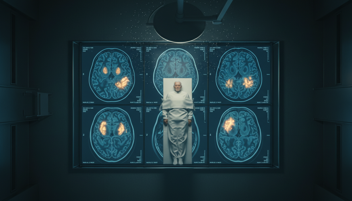
For Doctors in a Hurry
- This study aimed to determine if peri-ictal MRI abnormalities predict future unprovoked seizures after a first episode of status epilepticus.
- Researchers analyzed a prospective database of 135 adults without prior epilepsy who underwent MRI within 48 hours of the event.
- At 4 years, cumulative seizure probability was 61% for patients with DWI/FLAIR abnormalities versus 36% for those without such findings.
- The authors concluded that DWI/FLAIR abnormalities were independently associated with an increased probability of subsequent unprovoked seizures after status epilepticus.
- These specific MRI findings may help guide long-term treatment decisions by identifying patients at high risk for developing post-SE epilepsy.
From Acute Event to Chronic Disease: Forecasting Epilepsy After Status Epilepticus
Status epilepticus (SE) represents a critical neurologic emergency that necessitates rapid stabilization to mitigate the risk of permanent neurologic sequelae [1]. For the practicing clinician, a primary challenge following the resolution of the acute event is determining which patients will transition to chronic epilepsy, defined as an enduring biological predisposition to generate unprovoked seizures [2]. This prognostic uncertainty often complicates the decision to initiate long-term antiseizure medication, particularly in patients presenting with a first, or de novo, episode of status epilepticus, a clinical dilemma frequently encountered in the management of post-stroke seizures [3]. Identifying reliable, early biomarkers to stratify this risk is essential for optimizing long-term care [4]. A recent study investigated whether acute, transient findings on brain MRI performed shortly after the event can provide clinicians with a precise estimate of a patient's future seizure risk.
Imaging the Post-Ictal Brain: Study Design and Modalities
The researchers utilized a prospective database from the Christian Doppler University Hospital in Salzburg, Austria, to analyze a cohort of 135 adults who experienced their first episode of nonhypoxic status epilepticus. To ensure the findings applied specifically to de novo cases, the study excluded any patients with a prior history of epilepsy. The study population had a median age of 70 years (interquartile range 58 to 80 years), and 55% of the participants were female, reflecting a common clinical demographic where the decision to start lifelong medication carries significant implications for quality of life and drug-drug interactions. To capture acute neurobiological changes, all patients underwent MRI within 48 hours of their status epilepticus diagnosis.
Quantifying the Risk: DWI and FLAIR as Prognostic Markers
The study systematically evaluated peri-ictal MRI abnormalities (PMA), which are transient brain changes occurring around the time of a seizure, using three specific imaging sequences. These included diffusion-weighted imaging (DWI), a sequence sensitive to the restricted movement of water that identifies acute cytotoxic edema; T2-weighted fluid-attenuated inversion recovery (FLAIR), which suppresses cerebrospinal fluid signal to highlight parenchymal edema; and arterial spin labeling (ASL), a non-contrast perfusion method that uses magnetically labeled blood to measure regional cerebral blood flow. Over a median follow-up of 23 months (interquartile range 9 to 39 months), 43 patients (32%) experienced at least one unprovoked seizure. The presence of abnormalities on diffusion-weighted imaging or fluid-attenuated inversion recovery was associated with a cumulative seizure probability of 34% (95% CI 17 to 47) at 1 year and 61% (95% CI 37 to 75) at 4 years. This 61% risk is clinically significant as it meets the diagnostic threshold for epilepsy after a single event, suggesting that these imaging markers can justify the initiation of long-term antiseizure therapy. In contrast, patients with hyperperfusion on arterial spin labeling had a much lower cumulative seizure risk of 0% at 1 year and 13% (95% CI 0 to 29) at 4 years, while those without any peri-ictal MRI abnormalities had a risk of 25% (95% CI 14 to 34) at 1 year and 36% (95% CI 19 to 49) at 4 years.
Clinical and Electrographic Predictors of Recurrence
Beyond imaging, the clinical characteristics of the initial status epilepticus episode provided vital prognostic data. The researchers found that treatment refractoriness during the acute event was associated with a higher risk of subsequent seizures (odds ratio 2.93, 95% CI 1.23 to 6.98, p = 0.02). Additionally, a longer duration of status epilepticus increased the likelihood of recurrence (odds ratio 1.003, 95% CI 1.001 to 1.004, p = 0.02), suggesting that the cumulative burden of seizure activity may lower the seizure threshold over time. Electrographic findings also played a role: the presence of lateralized periodic discharges (repetitive, rhythmic electrographic patterns occurring over a single hemisphere that indicate focal cortical irritability) on ictal electroencephalogram was associated with a higher seizure risk (odds ratio 3.59, 95% CI 1.37 to 9.83, p = 0.005). These findings allow clinicians to integrate bedside clinical data with imaging to refine their prognostic estimates.
Independent Risk Factors and Clinical Application
To determine which factors most reliably predicted outcomes, the researchers employed a multivariable analysis, a statistical method that adjusts for multiple variables simultaneously to isolate the independent effect of each factor. This analysis confirmed that peri-ictal MRI abnormalities on diffusion-weighted imaging or fluid-attenuated inversion recovery were independently associated with increased seizure probability (log-odds 1.23, 95% CI 0.13 to 2.33, p = 0.03). The only other independent predictor was a remote etiology (log-odds 1.85, 95% CI 0.45 to 3.25, p = 0.009), which refers to a pre-existing brain lesion, such as a prior stroke or traumatic brain injury, that serves as a substrate for epilepsy. For the practicing physician, these results provide a clear framework: the presence of acute structural changes on MRI or a known prior brain injury significantly elevates the risk of chronic epilepsy following a first episode of status epilepticus. These markers can help guide long-term treatment decisions, identifying patients who may benefit most from early and sustained antiseizure intervention.
References
1. Shorvon S, Baulac M, Cross JH, Trinka E, Walker MC, Affairs FTTOSEOTICFE. The drug treatment of status epilepticus in Europe: Consensus document from a workshop at the first London Colloquium on Status Epilepticus. Epilepsia. 2008. doi:10.1111/j.1528-1167.2008.01706_3.x
2. Fisher RS, Boas WVE, Blume WT, et al. Epileptic Seizures and Epilepsy: Definitions Proposed by the International League Against Epilepsy (ILAE) and the International Bureau for Epilepsy (IBE). Epilepsia. 2005. doi:10.1111/j.0013-9580.2005.66104.x
3. Ravikumar A, Bhoopathy RM, Laha A, Mani R. Post – Stroke Seizures: A Comprehensive Review of Epidemiology, Pathophysiology, Risk Factors, Clinical Spectrum and Outcomes. Medical Research Archives. 2024. doi:10.18103/mra.v12i10.5768
4. Thaele A, Barba L, Abu-Rumeileh S, Foschi M, Otto M. Neurofilament light chain and glial fibrillary acidic protein as diagnostic and prognostic biomarkers in epileptic seizures and epilepsy: A systematic review.. Epilepsy & Behavior. 2025. doi:10.1016/j.yebeh.2025.110321