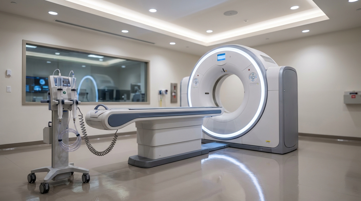
For Doctors in a Hurry
- Researchers investigated whether photon counting detector CT could maintain image quality for TAVR planning while using significantly reduced contrast media volumes.
- This retrospective study compared 162 body mass index matched examinations using either 80 milliliters or 50 milliliters of contrast media.
- Low energy reconstructions at 50 kiloelectron volts increased the contrast to noise ratio by 52.73 percent compared to standard protocols (p < 0.001).
- The authors concluded that photon counting technology preserves diagnostic efficacy for TAVR planning even when contrast agent dosing is minimized.
- This protocol offers a practical method to protect patients with renal impairment and mitigate environmental risks from iodinated contrast media.
Optimizing Pre-Interventional Imaging in Aortic Stenosis
The management of severe aortic stenosis relies heavily on transcatheter aortic valve replacement, a procedure requiring meticulous pre-interventional planning to ensure accurate valve sizing and vascular access assessment. Current clinical guidelines emphasize the necessity of comprehensive screening for coronary artery disease and structural abnormalities before these valvular interventions [1, 2]. While computed tomography is the established standard for this planning, the required bolus of iodinated contrast poses significant risks to a patient population frequently characterized by advanced age and comorbid renal dysfunction [3]. Furthermore, the prognostic importance of identifying myocardial fibrosis through advanced imaging highlights the growing demand for high-quality cardiac assessment in these high-risk patients [4, 5]. A recent study evaluates how photon-counting detector computed tomography (a technology that measures individual X-ray photons to improve spatial resolution and reduce noise) allows clinicians to significantly decrease contrast volumes while maintaining the diagnostic integrity of these essential scans.
Comparing Standard and Reduced Contrast Protocols
The researchers conducted a retrospective monocentric study involving 162 body mass index-matched examinations to evaluate the efficacy of reduced contrast protocols for transcatheter aortic valve reconstruction planning. This investigation utilized photon-counting detector computed tomography, an advanced imaging modality that counts individual X-ray photons to improve spatial resolution and reduce image noise, potentially allowing for lower iodine concentrations without sacrificing clarity. By matching patients based on body mass index, the authors ensured that differences in image quality were attributable to the contrast protocol and detector technology rather than variations in patient habitus. The study cohort was divided into two distinct groups to compare imaging outcomes. The standard contrast dose group consisted of 81 examinations using 80 mL of Iohexol 300 mg/mL. In comparison, the reduced contrast dose group included 81 examinations receiving only 50 mL of Iohexol 300 mg/mL, representing a 37.5 percent reduction in contrast volume. For clinicians managing patients with borderline renal function or those at risk for contrast-induced nephropathy, the transition from an 80 mL bolus to a 50 mL bolus provides a significant safety margin. This protocol adjustment addresses ongoing clinical concerns regarding iodine-related adverse events and the logistical challenges of recurring contrast media supply shortages, all while maintaining the precise anatomical mapping required for successful transcatheter valve placement.
Virtual Monoenergetic Reconstruction Techniques
To mitigate the loss of signal inherent in a 37.5 percent reduction of iodine, the researchers utilized virtual monoenergetic reconstructions, a post-processing technique that simulates images as if they were acquired at a single, specific X-ray energy level to enhance the attenuation of iodine. For the reduced contrast dose group, the authors generated virtual monoenergetic reconstructions at 70 keV, 60 keV, and 50 keV. These specific energy levels were selected to determine which setting best compensated for the lower contrast volume. These reconstructions were then compared with polyenergetic images, which are conventional computed tomography reconstructions that use the full spectrum of X-ray energies, to evaluate whether the specialized post-processing could restore the diagnostic clarity lost by using only 50 mL of contrast. The quantitative evaluation of image quality relied on the precise placement of regions-of-interest, defined areas on the scan used to measure tissue density and noise, across several critical anatomical landmarks. The researchers placed these regions-of-interest in the abdominal aorta, the iliac bifurcation, the femoral artery, the left ventricle, and the trapezius muscles. By measuring the attenuation and standard deviation within these zones, the team calculated the signal-to-noise ratio and the contrast-to-noise ratio for each reconstruction. This objective assessment allowed for a direct comparison between the standard 80 mL protocol and the various virtual monoenergetic reconstructions of the 50 mL protocol, ensuring that the vascular access and valve anatomy remained sufficiently visible for surgical planning.
Quantitative Gains in Signal and Contrast
The initial quantitative analysis revealed that reducing contrast media volume without specialized post-processing led to a measurable decline in image clarity. When comparing the reduced contrast dose group to the standard dose group using conventional reconstructions, the averaged signal-to-noise ratio decreased by 8.71 percent (p < 0.001). Similarly, the averaged contrast-to-noise ratio decreased by 16.78 percent (p < 0.001) in the reduced contrast cohort. These metrics represent the balance between the desired signal from the iodinated blood and the background noise that can obscure fine anatomical details, suggesting that a simple reduction in iodine volume without technical compensation could compromise the precision required for transcatheter aortic valve replacement planning. To counteract this degradation, the researchers applied low-energy virtual monoenergetic reconstructions, which significantly amplified the iodine signal. The use of virtual monoenergetic reconstructions at 50 keV increased the signal-to-noise ratio by 44.10 percent (p < 0.001) and the contrast-to-noise ratio by 52.73 percent (p < 0.001) when compared to the standard contrast dose protocol. This enhancement was particularly evident in the ascending aorta, a critical region for assessing valve morphology and annular dimensions. In this vessel, the signal-to-noise ratio increased from 19.80 ± 6.24 in the standard dose group to 35.78 ± 13.20 in the reduced dose 50 keV reconstruction group. The contrast-to-noise ratio in the ascending aorta also rose from 18.84 ± 7.78 in the standard dose group to 29.77 ± 16.70 in the 50 keV group, demonstrating that photon-counting detector technology can achieve superior image metrics even while using 37.5 percent less contrast media.
Clinical Utility and Diagnostic Integrity
Beyond quantitative metrics, the researchers conducted a qualitative assessment to ensure that the images met the rigorous standards required for surgical planning. Diagnostic quality and contrast were assessed qualitatively on a visual grading scale of 1 (non-diagnostic) to 5 (excellent). This subjective evaluation is critical for clinicians who must rely on these images to identify subtle anatomical landmarks and vascular pathologies. In the standard contrast dose group, which received 80 mL of Iohexol, the median contrast intensity was 5, representing excellent image quality. When the contrast volume was reduced to 50 mL without specialized post-processing, the median contrast intensity was 4 for the reduced contrast dose conventional reconstruction group. While still diagnostic, this decrease indicates a perceptible loss in opacification that could affect the confidence of a reading radiologist or interventionalist. The application of virtual monoenergetic reconstructions at 50 keV effectively bridged this gap. For the cohort receiving only 50 mL of contrast, the median contrast intensity was 5 for the reduced contrast dose 50 keV reconstruction group, matching the score of the full-dose protocol. The study concludes that diagnostic efficacy for transcatheter aortic valve replacement planning is preserved with minimized contrast dosing using photon-counting detector computed tomography. For the practicing clinician, these findings suggest that a substantial reduction in iodine load does not come at the cost of diagnostic certainty. This protocol offers a practical strategy to mitigate the risk of contrast-induced nephropathy in patients with renal impairment and helps manage institutional resources during periods of contrast media supply shortages.
References
1. Hosseini K, Sahzabi RY, Nasrollahizadeh A, et al. Diagnostic accuracy of CCTA versus ICA for detecting coronary artery disease in TAVI patients: a systematic review and meta-analysis. European Heart Journal. 2025. doi:10.1093/eurheartj/ehaf784.1687
2. Diller G, Gerwing M, Grazioli SB, et al. Utility of Coronary Computed Tomography Angiography in Patients Undergoing Transcatheter Aortic Valve Implantation: A Meta-Analysis and Meta-Regression Based on Published Data from 7458 Patients. Journal of Clinical Medicine. 2024. doi:10.3390/jcm13020631
3. Langenbach I, Langenbach MC, Mayrhofer T, et al. Reduction of contrast medium for transcatheter aortic valve replacement planning using a spectral detector CT: a prospective clinical trial. European Radiology. 2023. doi:10.1007/s00330-023-10403-x
4. Faggiano A, Gherbesi E, Carugo S, et al. Prognostic value of myocardial computed tomography–derived extracellular volume in severe aortic stenosis requiring aortic valve replacement: a systematic review and meta-analysis. European Heart Journal - Cardiovascular Imaging. 2025. doi:10.1093/ehjci/jeae324
5. Kirigaya J, Kato S, Matsushita K, Horita N, Utsunomiya D, Hibi K. Prognostic value of computed tomography-derived myocardial extracellular volume in aortic stenosis: a meta-analysis of all-cause mortality and heart failure hospitalization. European Heart Journal Open. 2024. doi:10.1093/ehjopen/oeaf007