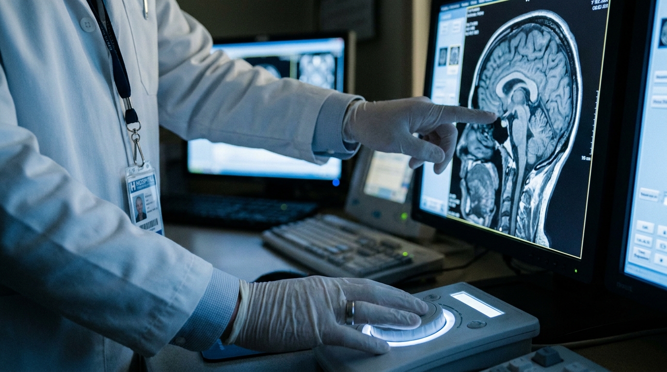
For Doctors in a Hurry
- Clinicians lack reliable tools to predict which oligometastatic patients will respond to ablative radiotherapy before initiating treatment.
- This scoping review analyzed 29 studies involving 3,946 patients to evaluate radiomics (extracting quantitative data from medical images).
- Models showed variable performance with area under the curve values between 0.69 and 0.95 across mostly brain metastases.
- The researchers concluded that while predictive performance is high, methodological heterogeneity and limited validation currently hinder clinical translation.
- Future multicenter prospective trials must standardize protocols before these models can help physicians identify patients unlikely to benefit.
Oligometastatic disease is increasingly recognized as a distinct intermediate state of cancer between localized and widespread metastatic forms (17). Stereotactic ablative radiotherapy has emerged as a standard intervention for these patients, with prospective data indicating improvements in both progression-free and overall survival (8, 13). However, the clinical presentation of this disease is highly heterogeneous, encompassing de novo, oligoprogressive, and induced states that may respond differently to local therapy (1, 2). While conventional prognostic factors such as tumor diameter and primary histology provide some guidance, they often lack the precision needed to predict the biological response of individual lesions to high-dose radiation (8, 12). A new systematic analysis now evaluates whether advanced computational analysis of imaging data can bridge this gap in clinical decision-making.
Analysis of Current Evidence and Imaging Modalities
Oligometastatic disease represents an intermediate stage of cancer, situated between localized disease and widespread systemic metastasis, and is frequently managed through surgical intervention or ablative radiotherapy (ART). To evaluate the current utility of advanced imaging analytics in this clinical setting, researchers conducted a scoping review following the Preferred Reporting Items for Systematic Reviews and Meta-Analyses (PRISMA) extension for Scoping Reviews guidelines. The investigators performed a systematic search across PubMed, Web of Science, Scopus, Embase, Cochrane, and Google Scholar, which initially identified 9463 records. After rigorous screening, the final analysis included 29 studies involving a total of 3946 patients, aiming to systematically summarize the evidence regarding radiomics (the extraction of large volumes of quantitative data from medical images) and its role in predicting clinical response to ART.
The review specifically examined how machine learning and deep learning (computational models that learn patterns directly from data) could be integrated with radiomics to refine treatment predictions. A significant majority of the included research relied on MRI-derived features to build these predictive models. Furthermore, the evidence base is currently heavily weighted toward intracranial applications, as 24 of the 29 included studies focused specifically on brain metastases. This concentration of data suggests that while radiomics shows potential for personalizing ablative radiotherapy, the current evidence is primarily centered on neurological sites rather than extracranial oligometastatic lesions.
Predictive Accuracy of Radiomics and Deep Learning
The analysis of the 29 included studies revealed that radiomics-based models demonstrated variable predictive performance, with an area under the curve (AUC) ranging from 0.69 to 0.95. This metric, which evaluates the ability of a diagnostic model to distinguish between patients who respond to treatment and those who do not, suggests that standard radiomic features can provide meaningful prognostic information. However, the highest levels of accuracy were observed in studies utilizing deep learning models, which are a subset of machine learning that uses multi-layered neural networks to automatically extract complex features from imaging data. These deep learning models achieved the highest accuracies with an AUC between 0.85 and 1.00, indicating a superior capacity for pattern recognition compared to traditional radiomic approaches.
From a clinical perspective, these findings suggest that radiomics-based models show significant potential for identifying patients who are unlikely to benefit from ablative radiotherapy. For the practicing clinician, this capability is particularly relevant for treatment stratification; by identifying non-responders before the initiation of therapy, physicians may be able to spare patients from the toxicity and costs associated with ineffective ablative radiotherapy. While the current evidence is strongest for brain metastases, the high predictive accuracy of these computational models provides a foundation for more personalized oncology workflows, provided that these tools undergo further validation in prospective, multicenter clinical settings.
Methodological Quality and Reporting Standards
To determine the reliability of the current evidence base, the researchers evaluated the rigor of the 29 included studies using standardized assessment tools designed specifically for high-dimensional imaging data. The authors employed the Radiomics Quality Score (RQS), a metric that assesses the scientific integrity and clinical relevance of radiomics workflows, alongside the METhodological RadiomICs Score (METRICS), which evaluates the technical validity of the modeling process. Furthermore, reporting transparency was evaluated using the CheckList for EvaluAtion of Radiomics research (CLEAR), a tool designed to ensure that studies provide sufficient detail for other researchers to reproduce the findings and for clinicians to interpret the results accurately.
The analysis revealed that the methodological quality of the included studies was moderate, indicating that while the findings are statistically significant, the evidence base requires further refinement before widespread clinical adoption. Specifically, the mean Radiomics Quality Score (RQS) across the studies was 13, a score that reflects common limitations in retrospective study designs and a lack of external validation cohorts. Additionally, the METRICS scores for the studies ranged from 64.2% to 78%, highlighting a degree of variability in how researchers handled image preprocessing and feature selection. For the practicing clinician, these scores serve as a reminder that while the predictive accuracy of these models is high, the underlying evidence is currently constrained by methodological heterogeneity, necessitating a cautious approach to clinical implementation until more standardized, prospective data become available.
Barriers to Clinical Translation and Future Directions
Despite the high predictive accuracy observed in deep learning models, clinical implementation of these radiomics models remains limited, particularly for patients presenting with extracranial metastases. The scoping review of 29 studies involving 3,946 patients highlighted that substantial methodological heterogeneity and limited validation are the primary factors currently hindering the translation of these computational tools into routine oncology practice. While the evidence base for brain metastases is more robust, the lack of standardized data for other anatomical sites prevents a broader application of radiomics in the management of the intermediate state of cancer known as oligometastatic disease.
To bridge the gap between research and clinical utility, the authors emphasize that future research must transition toward multicenter, prospective studies utilizing standardized protocols to ensure the reproducibility of imaging features across different institutions and scanner types. Furthermore, the next generation of predictive tools should move beyond imaging features alone by incorporating clinical and dosimetric data, which provides critical information regarding the spatial distribution and absorbed dose of radiation within the target tissue. Integrating these variables is essential for developing comprehensive models that can reliably guide personalized treatment decisions and identify which patients are most likely to benefit from ablative radiotherapy.