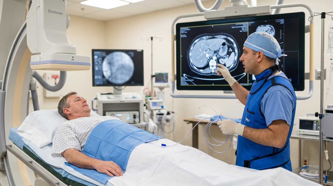
For Doctors in a Hurry
- Clinicians lack reliable tools to select patients with unresectable colorectal liver metastases who will benefit from transarterial chemoembolization.
- Researchers analyzed 76 patients with 176 lesions from a multicenter registry to develop machine learning models based on radiomic features.
- Baseline intensity models predicted survival with an area under the curve of 0.79 (95% CI, 0.57-0.95; p=0.011).
- The study concluded that radiomics-based machine learning effectively stratifies survival risk and identifies treatment response in patients receiving irinotecan-based chemoembolization.
- These models may improve patient selection and personalized therapy by identifying individuals most likely to benefit from locoregional interventions.
Colorectal cancer frequently metastasizes to the liver, with only approximately 20% of patients presenting with resectable disease at the time of diagnosis [1]. For the majority with unresectable colorectal liver metastases, regional therapies such as drug-eluting bead transarterial chemoembolization (TACE, a procedure where microspheres deliver concentrated chemotherapy directly to the tumor arterial supply) using irinotecan have emerged as important options, particularly in the second-line setting [2, 3]. While these intra-arterial interventions can improve median progression-free survival (the time a patient lives without the disease worsening) to 6.7 months compared to 3.8 months with systemic therapy alone (p = 0.009), clinical outcomes remain highly variable [4, 5]. Identifying which individuals will derive a meaningful survival benefit remains a significant challenge for multidisciplinary oncology teams [1, 6]. A recent analysis of the prospective CIREL registry trial now evaluates whether radiomics (the extraction of quantitative data from medical images to uncover disease characteristics invisible to the human eye) can provide the prognostic precision needed to guide treatment, specifically using a baseline intensity algorithm that predicted overall survival with an Area Under the Curve (AUC) of 0.79 (95% CI = 0.57 to 0.95) [7].
Machine Learning Analysis of the CIREL Registry
The researchers conducted a retrospective analysis of data from the prospective CIREL registry trial, focusing on a cohort of 76 patients who presented with unresectable colorectal liver metastases. Within this study population, the investigators evaluated a total of 176 lesions to determine the efficacy of various imaging markers in predicting clinical outcomes. The demographic profile of the final study population showed a median age of 66 years, with an interquartile range of 59 to 71 years. Of these participants, 67.1% (n = 51) identified as male, providing a representative sample of the typical patient population undergoing locoregional therapy for metastatic colorectal disease. To develop a robust predictive framework, the study tested three distinct categories of imaging markers: general radiomics, intensity-based features, and lesion volume. General radiomics involves the extraction of high-dimensional data regarding tumor shape and texture, while intensity-based features focus on the distribution of voxel gray-level values within the tumor. For each of these markers, the researchers derived both baseline features and delta features, which represent the specific difference in feature vector values between the initial baseline scan and the first follow-up imaging. This longitudinal approach allowed the team to quantify how the internal characteristics of the tumor shifted in response to the initial treatment phase. The analysis utilized genetic/evolutionary machine learning models (algorithms that iteratively select the most predictive variables by mimicking the process of natural selection) to predict both overall survival and lesion-level response. To ensure the reliability of these models, the researchers employed a center-based split for training and validation. This rigorous method involves training the algorithm on data from specific medical centers and testing it on data from entirely different facilities, ensuring that the predictive power of the model remains consistent across varying clinical environments and imaging protocols.
Baseline Intensity as a Superior Predictor of Survival
The researchers evaluated the capacity of different imaging markers to predict overall survival following irinotecan-TACE, focusing on how pre-treatment characteristics correlate with long-term clinical outcomes. While clinicians often rely on tumor size to gauge prognosis, this study suggests that internal tumor characteristics are more informative for this specific patient population. Specifically, the investigators compared the predictive accuracy of baseline intensity features (which quantify the distribution of gray-level values within the tumor) against general radiomics and lesion volume. Upon external validation, the baseline intensity algorithm emerged as the only significant survival-prediction model, achieving an AUC of 0.79 (95% CI = 0.57 to 0.95; p = 0.011). This metric indicates a high level of predictive accuracy for intensity-based features on pre-treatment imaging. In contrast, baseline radiomics (which incorporates complex data regarding tumor shape and texture) achieved an AUC of 0.69 (95% CI = 0.47 to 0.86; p = 0.100). Even more striking was the poor performance of baseline volume, a standard clinical metric, which yielded an AUC of only 0.56 (95% CI = 0.37 to 0.74; p = 0.574). These findings suggest that simple volumetric measurements are insufficient for stratifying survival risk in patients with unresectable colorectal liver metastases, whereas the distribution of voxel intensities provides a more reliable prognostic signal. For the practicing physician, this means that looking beyond simple tumor diameter on a scan could eventually help identify which patients are truly candidates for transarterial chemoembolization, sparing unlikely responders from unnecessary procedural risks.
Stratifying Patient Risk and Predicting Lesion Response
The clinical utility of these machine learning models extends beyond simple prediction to the active stratification of patient outcomes. By analyzing the extracted imaging data, the radiomic models successfully stratified patients into distinct low-risk and high-risk groups for overall survival. This differentiation resulted in a statistically significant gap in clinical outcomes, as low-risk patients demonstrated a median survival of 696 days compared to 453 days for those in the high-risk group (log-rank p = 0.0267). For the practicing oncologist, this nearly eight-month difference in median survival provides a quantitative basis for discussing prognosis and setting realistic expectations for patients undergoing irinotecan-TACE for metastatic liver disease. Beyond baseline predictions, the researchers evaluated how longitudinal changes in tumor characteristics could track treatment efficacy. The study found that lesion-level response was best identified by delta radiomics (the calculated difference in imaging features between the baseline scan and the first follow-up), achieving an AUC of 0.83 (95% CI = 0.63 to 0.97; p = 0.008). This suggests that the evolving texture and intensity patterns within a lesion are more sensitive indicators of treatment response than traditional diameter-based assessments. The investigators also explored whether combining imaging data with clinical laboratory variables could further refine these predictions. Integrating imaging features with laboratory variables improved the assessment of lesion-level response, reaching an AUC of 0.86 (95% CI = 0.66 to 0.99; p = 0.006). However, the addition of these laboratory variables did not enhance the prediction of overall survival. This distinction is critical for clinical workflow, as it suggests that while blood-based biomarkers may help confirm whether a specific lesion is responding to chemoembolization, they do not necessarily add prognostic value to the survival data already provided by baseline imaging intensity. Ultimately, these tools may allow clinicians to personalize therapy by identifying patients likely to benefit from irinotecan-TACE while sparing non-responders from the toxicity of futile interventions.
References
1. Karanicolas P, Beecroft JR, Cosby R, et al. Regional Therapies for Colorectal Liver Metastases: Systematic Review and Clinical Practice Guideline.. Clinical colorectal cancer. 2021. doi:10.1016/j.clcc.2020.09.008
2. Richardson AJ, Laurence JM, Lam VWT. Transarterial chemoembolization with irinotecan beads in the treatment of colorectal liver metastases: systematic review.. Journal of vascular and interventional radiology : JVIR. 2013. doi:10.1016/j.jvir.2013.05.055
3. Zhao JJ, Tan E, Sultana R, et al. Intra-Arterial Therapy for Unresectable Colorectal Liver Metastases: A Meta-Analysis.. Journal of vascular and interventional radiology : JVIR. 2021. doi:10.1016/j.jvir.2021.05.032
4. Sugumar K, Stitzel H, Wu V, et al. Outcomes of Hepatic Artery-Based Therapies and Systemic Multiagent Chemotherapy in Unresectable Colorectal Liver Metastases: A Systematic Review and Meta-analysis.. Annals of surgical oncology. 2024. doi:10.1245/s10434-024-15187-y
5. Liu Y, Chang W, Zhou B, et al. Conventional transarterial chemoembolization combined with systemic therapy versus systemic therapy alone as second-line treatment for unresectable colorectal liver metastases: randomized clinical trial.. The British journal of surgery. 2021. doi:10.1093/bjs/znaa155
6. Levy J, Zuckerman J, Garfinkle R, et al. Intra-arterial therapies for unresectable and chemorefractory colorectal cancer liver metastases: a systematic review and meta-analysis.. HPB. 2018. doi:10.1016/j.hpb.2018.04.001
7. Bodalal Z, Ferradás FJM, Maxouri O, et al. Radiomics-based outcome prediction for irinotecan-TACE in colorectal liver metastases: advanced analysis from the prospective CIREL trial.. European radiology. 2026. doi:10.1007/s00330-026-12573-w