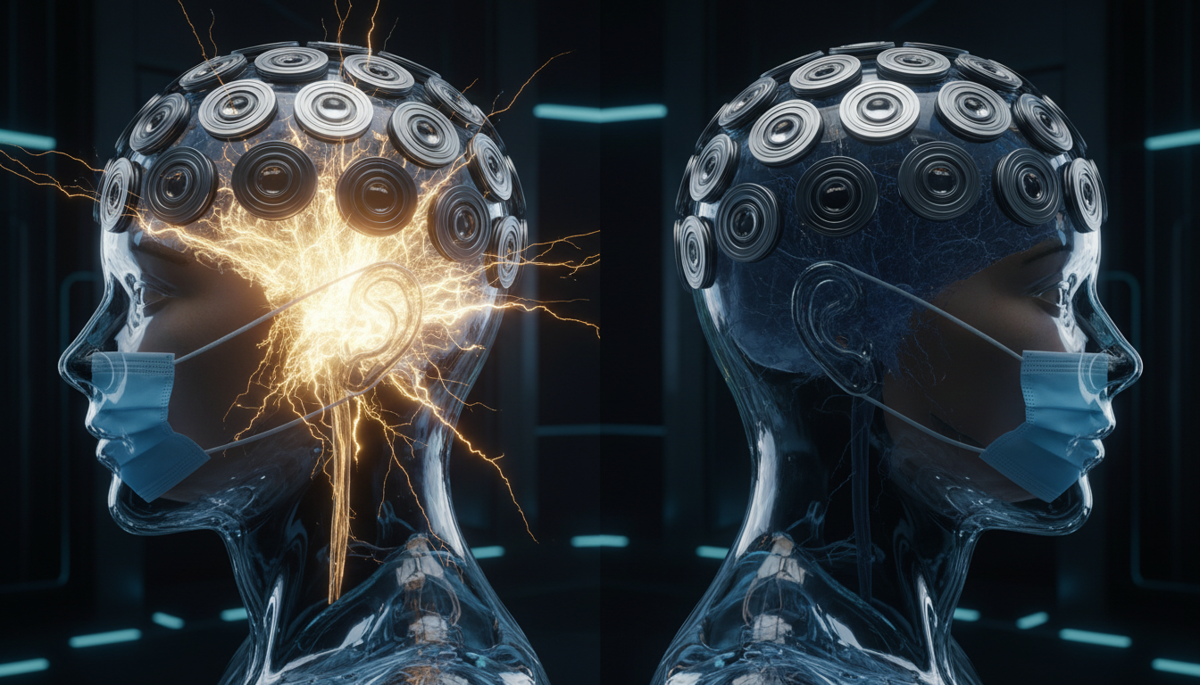
For Doctors in a Hurry
- Researchers investigated whether patients with schizophrenia exhibit abnormal neural responses when internal visual predictions conflict with new sensory evidence.
- The study used magnetoencephalography to measure brain activity in 18 patients with schizophrenia and 18 healthy controls viewing masked and unmasked faces.
- Healthy controls demonstrated a significant left temporal sensor response between 234 and 511 milliseconds involving the insular cortex, whereas patients with schizophrenia showed none.
- The authors concluded that patients with schizophrenia lack robust neural mismatch responses when internal facial representations meet newly revealed sensory information.
- These findings support predictive processing theories of reduced perceptual inference precision in schizophrenia, offering clinicians a mechanistic explanation for altered sensory perception.
The Predictive Brain and Perceptual Deficits in Schizophrenia
Schizophrenia is characterized by profound disruptions in perception and cognition, often manifesting as hallucinations, delusions, and severe social withdrawal. A growing body of evidence suggests these symptoms may stem from deficits in predictive processing, where the brain struggles to accurately integrate internal expectations with incoming sensory data, a phenomenon frequently studied using mismatch negativity paradigms [1, 2]. Furthermore, patients consistently demonstrate specific impairments in facial recognition and social cognition, which correlate with altered electrophysiological responses and disrupted cortico-limbic connectivity [3, 4]. While traditional mismatch studies focus on violations of external patterns, understanding how the brain reacts when its own internal predictions fail remains a critical gap in the pathophysiology of psychosis. A new study now offers fresh insights into this mechanism by examining how the brain processes the sudden revelation of hidden facial features.
Probing Internal Predictions with Magnetoencephalography
Predictive-processing accounts have become increasingly influential in understanding the pathophysiology of schizophrenia. Within this framework, researchers frequently utilize mismatch negativity paradigms, which are established tests that measure the brain's automatic electrical response to unexpected stimuli. Clinically, these paradigms help physicians understand why patients with psychosis often struggle to filter and interpret incoming sensory information. However, most mismatch negativity designs define a mismatch as a violation of externally defined regularities, such as a sudden change in a repetitive sequence of sounds or images. To expand on this traditional model, the researchers utilized magnetoencephalography (a noninvasive neuroimaging technique that maps brain activity by recording magnetic fields produced by electrical currents) to probe a complementary form of mismatch. This specific reaction, known as an inferred-sensory mismatch, arises when an internally inferred representation of an occluded face is confronted with newly available sensory evidence after unmasking.
During the experiment, participants first viewed masked faces, followed immediately by the same faces without masks. The researchers used two types of stimuli (individual faces and average composite faces) to determine whether the brain responds differently to specific personal features compared to generalized facial structures. To investigate these neural dynamics, the authors analyzed data from 18 individuals with schizophrenia and 18 healthy controls. The data were evaluated using spatiotemporal cluster tests, a statistical approach that groups adjacent data points in time and physical brain space to identify significant patterns of neural activity while controlling for false positive rates. This design allowed the investigators to precisely track how the brain updates its internal models the exact moment hidden facial features are revealed.
Absence of Neural Mismatch in Schizophrenia
The findings revealed distinct neurological differences between the two cohorts when hidden facial features were suddenly revealed. Among healthy controls, mask removal from individual faces elicited a significant cluster of activity at the left temporal sensors. This burst of neural processing occurred between 234 and 511 milliseconds after the mask was removed. Furthermore, source estimates for this significant cluster in healthy controls suggested the involvement of the left insular cortex, a brain region heavily implicated in sensory processing and self-awareness. Ultimately, healthy controls demonstrated a reliable neural response to the unmasking of individual faces, consistent with a mismatch between an internally inferred representation and newly revealed sensory information. Interestingly, this effect was highly specific to distinct personal features, as no significant clusters were observed for average faces in the healthy control group.
The neurological reaction in patients with psychosis was markedly different. In the schizophrenia group, no significant mismatch responses were observed for either individual or average faces. Overall, no robust mismatch-related response was detected in the schizophrenia cohort. For practicing physicians, this absence of a neural signal provides a biological correlate for the cognitive symptoms often seen in the clinic. This pattern is compatible with predictive-processing accounts that emphasize a reduced precision of perceptual inference in schizophrenia. In practical terms, this means the brains of patients with schizophrenia may struggle to generate strong internal predictions or fail to register when those predictions conflict with actual sensory input. This fundamental deficit in updating internal models with new sensory evidence likely contributes to the perceptual distortions, delusions, and hallucinations that characterize the disorder, highlighting a potential neurobiological target for future therapeutic interventions.
References
1. Varvari I, Bolte L, Colli C, et al. Glutamatergic Modulation of Brain Function in Psychosis: A Systematic Review of Neuroimaging Studies.. Biological psychiatry. Cognitive neuroscience and neuroimaging. 2025. doi:10.1016/j.bpsc.2025.07.004
2. Rosburg T, Kreitschmann-Andermahr I. The effects of ketamine on the mismatch negativity (MMN) in humans - A meta-analysis.. Clinical neurophysiology : official journal of the International Federation of Clinical Neurophysiology. 2016. doi:10.1016/j.clinph.2015.10.062
3. Scala M, Dukart J, Simone GD, et al. Magnetoencephalographic signatures of facial emotion processing in schizophrenia and major depressive disorder: a systematic review.. Neuropsychobiology. 2026. doi:10.1159/000550903
4. Feuerriegel D, Churches O, Hofmann J, Keage HAD. The N170 and face perception in psychiatric and neurological disorders: A systematic review.. Clinical neurophysiology : official journal of the International Federation of Clinical Neurophysiology. 2015. doi:10.1016/j.clinph.2014.09.015