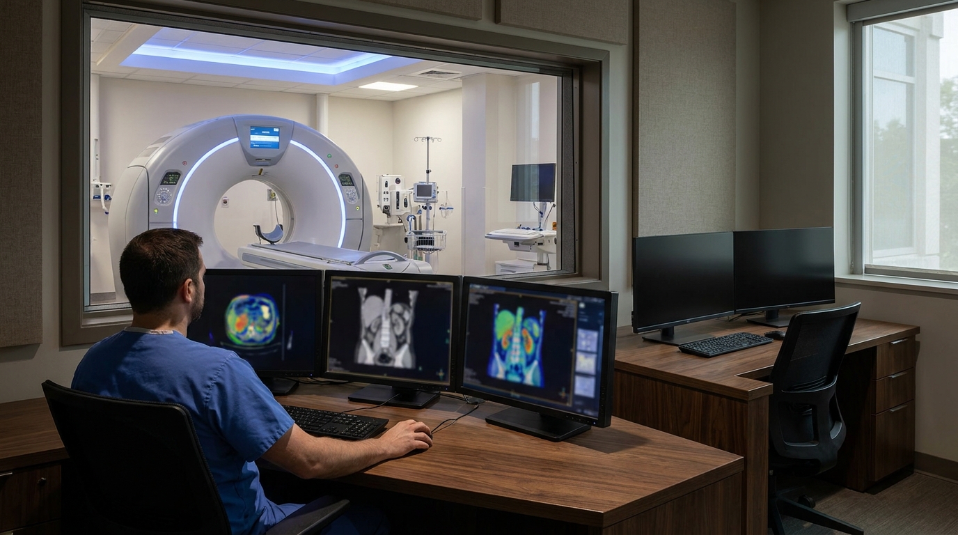
For Doctors in a Hurry
- Researchers investigated whether adding spectral reconstructions to portal-venous phase CT images improves the detection of acute bowel ischemia.
- This retrospective study evaluated 378 patients using dual-energy and photon-counting CT to compare blended images against supplemental spectral reconstructions.
- Adding spectral reconstructions increased diagnostic accuracy from 73 percent to 86 percent (p < 0.001) and sensitivity to 87 percent.
- The researchers concluded that spectral reconstructions significantly improve diagnostic performance and confidence for bowel ischemia across both CT imaging platforms.
- These findings suggest that spectral reconstructions may improve emergency abdominal CT interpretation without requiring additional radiation or acquisitions.
Refining the Diagnostic Approach to Acute Bowel Ischemia
Acute bowel ischemia remains a critical diagnostic challenge in the emergency department, where rapid identification of non-viable tissue is essential to prevent gangrene and reduce mortality [1, 2]. While conventional computed tomography serves as the primary workup tool, subtle perfusion deficits can be difficult to distinguish on standard blended images alone. To address this, dual-energy computed tomography utilizes iodine quantification (a method measuring contrast concentration within tissues) and virtual monoenergetic imaging (a reconstruction technique simulating specific energy levels to enhance contrast) to improve vascular visualization [3, 4, 5]. These spectral techniques have demonstrated utility in reducing contrast volumes and identifying gastrointestinal bleeding with high sensitivity [3, 6]. Highlighting this potential, a prior retrospective study of 50 patients found that adding iodine maps and virtual non-contrast images significantly increased diagnostic sensitivity from 77% to 89% (p = 0.02) and improved reader confidence among both residents and fellowship-trained radiologists [7].
Multireader Evaluation of Spectral Reconstruction Protocols
To evaluate how spectral computed tomography data influences the detection of acute bowel ischemia, researchers conducted a retrospective, single-center, multireader, crossover diagnostic accuracy study. The study cohort consisted of 378 patients with a mean age of 68.1 years (± 7.4 years), including 206 men. To establish a definitive diagnosis for comparison, the authors utilized a prespecified composite reference standard derived from surgical findings and index-hospitalization documentation. Within this patient group, 150 out of 378 individuals (39.7%) were confirmed as ischemia-positive, providing a robust dataset for assessing diagnostic performance across different imaging protocols.
The evaluation compared the diagnostic performance of portal venous blended computed tomography images alone (designated as image set A) against a supplemented protocol (image set B). This second protocol added three specific spectral reconstructions to the standard portal venous blended images: 40-keV virtual monoenergetic images, iodine maps, and virtual non-contrast images. The 40-keV virtual monoenergetic images are low-energy reconstructions that enhance iodine contrast to make subtle perfusion defects more visible to the interpreting radiologist. Iodine maps isolate the distribution of iodine within the tissue to quantify blood flow, while virtual non-contrast images mathematically remove iodine from the scan to simulate a pre-contrast acquisition. For the practicing clinician, this means radiologists can better differentiate between active contrast extravasation and pre-existing hyperdense material, potentially accelerating the time to definitive surgical intervention.
Quantifying Gains in Sensitivity and Specificity
The researchers utilized mixed-effects logistic models (statistical tools that account for both fixed effects like the imaging protocol and random effects like patient variability) to evaluate primary diagnostic metrics. They found that diagnostic accuracy increased from 73% (95% CI: 68-77) with blended images to 86% (95% CI: 82-89) with spectral reconstructions. This improvement was statistically robust, yielding an odds ratio for increased accuracy of 2.30 (95% CI: 1.70-3.10; p < 0.001). To further validate these findings across different clinical scenarios, the study employed receiver operating characteristic analyses using a multireader multicase framework, a statistical design that accounts for variability among both the interpreting radiologists and the individual patient cases.
The addition of spectral data significantly enhanced the detection of ischemic changes while simultaneously reducing false positives. Sensitivity increased from 75% (95% CI: 68-82) with blended images to 87% (95% CI: 81-92) with spectral reconstructions (p = 0.008), while specificity rose from 72% (95% CI: 65-78) to 86% (95% CI: 80-90) (p < 0.001). These gains were reflected in the mean Area Under the Curve, which improved by 0.16 (95% CI: 0.12-0.20; p < 0.001). In clinical terms, the Area Under the Curve represents the overall ability of the imaging test to correctly distinguish between patients with bowel ischemia and those without the condition, where a value of 1.0 represents a perfect test.
Beyond binary diagnostic metrics, the researchers assessed how spectral reconstructions influenced the subjective certainty of the interpreting physicians. Using cumulative link mixed models (statistical methods used to evaluate ordered categorical data), the authors analyzed 7-point suspicion scores assigned by the readers. The results indicated that adding spectral reconstructions improved diagnostic confidence for bowel ischemia detection, as evidenced by more definitive suspicion scores compared to those generated using portal venous blended images alone. For emergency physicians and surgeons, this translates to radiology reports that offer clearer, more definitive guidance when deciding whether to take a critically ill patient to the operating room.
The researchers evaluated the utility of spectral reconstructions across two distinct imaging technologies: dual-energy computed tomography (DECT) and photon-counting computed tomography (PCCT). While dual-energy computed tomography has been the established standard for spectral imaging, photon-counting computed tomography represents a newer detector technology that counts individual X-ray photons to improve spatial resolution and reduce image noise. Despite the technical differences in how these scanners acquire data, the study demonstrated that the diagnostic benefits of spectral reconstructions remained robust across both systems.
Statistical analysis confirmed that the improvements in diagnostic performance were not dependent on the specific hardware used. The researchers found that the diagnostic effects were similar on DECT and PCCT platforms, as indicated by an interaction p-value of 0.24. This lack of evidence for platform-specific effect modification suggests that clinicians can expect comparable gains in accuracy, sensitivity, and specificity regardless of whether their institution utilizes traditional dual-energy or newer photon-counting equipment.
From a clinical workflow perspective, these findings are particularly relevant because the spectral reconstructions are derived from a single portal venous acquisition. This allows for the generation of iodine maps and monoenergetic images without the need for additional scans or increased radiation exposure. Consequently, the use of these advanced reconstructions may improve emergency abdominal computed tomography interpretation for bowel ischemia without additional radiation to the patient. For the practicing physician, this means a higher diagnostic yield from a standard emergency scan, facilitating rapid surgical triage without compromising patient safety through increased X-ray dose.
References
1. Choi J, Yang S, Lee JH, et al. Maternal Bochdalek Hernia during Pregnancy: A Systematic Review of Case Reports. Diagnostics. 2021. doi:10.3390/diagnostics11071261
2. Koelemay MJ, Geelkerken RH, Kärkkäinen JM, et al. Editor's Choice – European Society for Vascular Surgery (ESVS) 2025 Clinical Practice Guidelines on the Management of Diseases of the Mesenteric and Renal Arteries and Veins. European Journal of Vascular and Endovascular Surgery. 2025. doi:10.1016/j.ejvs.2025.06.010
3. Guerrini S, Zanoni M, Sica C, et al. Dual-Energy CT as a Well-Established CT Modality to Reduce Contrast Media Amount: A Systematic Review from the Computed Tomography Subspecialty Section of the Italian Society of Radiology. Journal of Clinical Medicine. 2024. doi:10.3390/jcm13216345
4. Xu JJ, Taudorf M, Ulriksen P, et al. Gastrointestinal Applications of Iodine Quantification Using Dual-Energy CT: A Systematic Review. Diagnostics. 2020. doi:10.3390/diagnostics10100814
5. Albrecht MH, Vogl TJ, Martin SS, et al. Review of Clinical Applications for Virtual Monoenergetic Dual-Energy CT. Radiology. 2019. doi:10.1148/radiol.2019182297
6. Oberparleiter M, Breit H, Vosshenrich J, et al. Replacing true non-contrast imaging with DECT in GI bleeding demonstrates non-inferior diagnostic performance, reading time and confidence. European Radiology. 2025. doi:10.1007/s00330-025-12191-y
7. Oberparleiter M, Vosshenrich J, Breit H, et al. Dual-energy CT of acute bowel ischemia—influence on diagnostic accuracy and reader confidence. European Radiology. 2024. doi:10.1007/s00330-024-11217-1