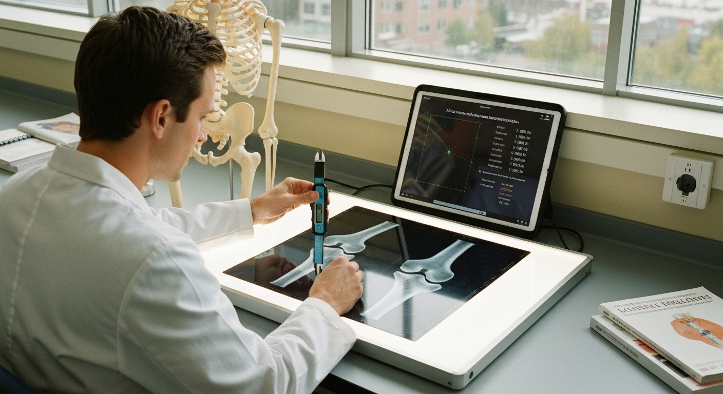
For Doctors in a Hurry
- Clinicians require efficient methods to reduce inter-reader variability and manual labor during musculoskeletal radiographic morphometric measurements.
- The researchers analyzed 600 standard radiographs and 240 images with orthopedic implants using a training-free artificial intelligence framework.
- Mean landmark matching error reached 2.15 millimeters with 40 reference radiographs, while measurement accuracy varied between 1.81 and 8.65 degrees.
- The authors concluded that this anatomy-agnostic framework achieves measurement accuracy often comparable to inter-reader agreement among radiologists.
- This tool may automate repetitive measurements to improve reproducibility, provided clinicians implement quality control for challenging cases.
Standardizing Morphometric Assessment in Musculoskeletal Imaging
Musculoskeletal disorders remain a primary driver of global disability, necessitating highly precise diagnostic and management strategies to optimize patient outcomes [1]. In clinical practice, the interpretation of medical images and the prediction of surgical success often rely on detailed morphometric measurements, yet these tasks are frequently hindered by high inter-reader variability and significant manual labor [2, 3]. While artificial intelligence has shown potential in detecting fractures and grading osteoarthritis, many existing models are limited by their narrow focus on specific joints or the requirement for massive, annotated datasets [1, 4]. Furthermore, the integration of these digital tools into routine workflows is often stalled by the black-box nature of complex algorithms and a lack of standardized validation [5, 6]. A new study now evaluates a generalist framework designed to automate these measurements across multiple anatomical regions using a universal matching approach.
Universal Landmark Matching Methodology
Current artificial intelligence solutions in radiology often require large annotated training datasets and are limited by narrow applications, typically focusing on a single joint or specific radiographic view. To address these constraints, the researchers validated a training-free artificial intelligence framework that automatically derives morphometric measurements across multiple anatomies and radiographic views. This system utilizes universal landmark matching, a method that identifies key anatomical points without the need for the extensive, site-specific data labeling that characterizes traditional machine learning models. By removing the requirement for thousands of hand-labeled images, the framework allows for a more flexible implementation across diverse clinical presentations. The core of this framework involves a pre-trained generalist dense-matching method, which is a technique that maps corresponding points between images without specific training on those images. By transferring landmarks from a small set of reference radiographs to unseen patient images, the system establishes a geometric foundation for analysis. Following this landmark transfer, the actual clinical measurements are derived in a post-processing step. While the framework eliminates the need for lengthy training phases, the researchers noted that clinically practical runtimes for this framework require Graphics Processing Unit (GPU) inference (the use of specialized high-speed processors) to ensure the computational speed necessary for real-time diagnostic workflows in a busy clinic. Because the framework is anatomy-agnostic, meaning it can be applied to various body parts without recalibration, it enables training-free morphometry across multiple regions, including the foot, knee, and shoulder. The study found that this approach yields measurement-dependent performance often comparable to inter-reader agreement between expert radiologists. Furthermore, the minimal setup required for the framework enables rapid adaptation to new anatomies and measurements. This allows clinicians to implement automated assessments for various musculoskeletal conditions without developing new, specialized algorithms for every clinical scenario, effectively bridging the gap between generalized AI and specific orthopedic diagnostic needs.
Validation Against Radiologist Standards
To evaluate the clinical utility of the universal landmark matching framework, the researchers conducted a retrospective study analyzing 600 standard radiographs of the foot, knee, and shoulder. This diverse dataset allowed for a robust assessment of the system across different anatomical structures and imaging planes. To establish a ground truth for comparison, the results generated by the artificial intelligence were compared with manual annotations and measurements performed by two expert radiologists. This comparison aimed to determine if the automated system could match the precision of human specialists in identifying key anatomical landmarks and calculating the resulting clinical angles. The accuracy of the system was primarily measured by the mean landmark matching error, which represents the distance between the AI-placed point and the expert-defined location. When the framework utilized only a single reference radiograph, the mean landmark matching error was 2.68 ± 2.70 mm. However, the researchers found that the system's precision is scalable; the mean landmark matching error improved to 2.15 ± 2.38 mm when using 40 reference radiographs. This data suggests that providing the model with a broader library of anatomical variations allows it to more accurately map landmarks on unseen patient images, reducing the margin of error toward a level acceptable for clinical decision-making. Beyond individual points, the study assessed the accuracy of specific clinical measurements derived from these landmarks. The results showed that measurement accuracy ranges from 1.81° for the I–II metatarsal angle to 8.65° for the congruence angle, reflecting the inherent complexity of different radiographic views. A critical finding for practicing clinicians is that increasing the number of reference images improved measurement accuracy and mostly approached inter-reader agreement. This indicates that with a sufficient reference set, the automated framework can produce morphometric data that is statistically similar to the variations seen between two human experts, potentially offering a more reproducible alternative for routine orthopedic assessments.
Accurate morphometric measurements are crucial for musculoskeletal radiography, as they provide the objective data necessary for surgical planning and the longitudinal monitoring of degenerative conditions. Despite their clinical importance, these assessments remain labor-intensive and prone to inter-reader variability, where the subjective placement of landmarks by different clinicians can lead to inconsistent diagnostic conclusions. To address these challenges, the researchers evaluated whether a generalist artificial intelligence framework could provide the reproducibility required for routine clinical practice without the need for extensive, anatomy-specific training sets. To evaluate the robustness of the system beyond ideal imaging conditions, the researchers constructed a cohort of 240 challenging radiographs containing orthopedic implants to stress-test the approach. This dataset included images where standard anatomical landmarks were potentially obscured or altered by metal hardware, such as plates, screws, or prosthetic components. The study found that performance is mixed on the challenging cohort containing orthopedic implants, demonstrating specific limitations and strengths of the universal landmark matching method. While the framework maintained some utility in these complex scenarios, the presence of non-biological materials introduced higher error rates than those seen in standard anatomy. These challenging cases highlight limitations that motivate the use of quality control and reference-set tuning for clinical deployment. Because the system relies on matching landmarks from a reference library to a new patient image, the researchers noted that the accuracy in post-operative cases depends heavily on having reference images that reflect similar surgical alterations. For the practicing clinician, these findings suggest that while the automated framework can significantly reduce manual workload, its integration into a professional workflow requires a supervised approach, particularly when managing patients with complex orthopedic histories or significant hardware.
References
1. Zanib A, Riaz F, Nazar S, Afaq E, Askari Z, Moeez S. Sub135 SYSTEMATIC REVIEW ON THE ROLE OF ARTIFICIAL INTELLIGENCE IN DIAGNOSING AND MANAGING MUSCULOSKELETAL DISORDERS AND INJURIES. 2025. doi:10.71000/v077b679
2. Longo UG, Marino M, Nicodemi G, et al. Artificial intelligence applications in the management of musculoskeletal disorders of the shoulder: A systematic review.. Journal of experimental orthopaedics. 2025. doi:10.1002/jeo2.70248
3. Droppelmann G, Rodríguez C, Jorquera C, Feijoo F. Artificial intelligence in diagnosing upper limb musculoskeletal disorders: a systematic review and meta-analysis of diagnostic tests.. EFORT open reviews. 2024. doi:10.1530/EOR-23-0174
4. Manzhalii E, Dekhtiar Y, Bannikov V, Girnyk G, Bavykin I. ARTIFICIAL INTELLIGENCE IN CLINICAL DIAGNOSTICS FOR EARLY DETECTION OF CHRONIC DISEASES: A SYSTEMATIC REVIEW.. Georgian medical news. 2026.
5. Xiang L, Gao Z, Yu P, et al. Explainable artificial intelligence for gait analysis: advances, pitfalls, and challenges - a systematic review.. Frontiers in bioengineering and biotechnology. 2025. doi:10.3389/fbioe.2025.1671344
6. Jayakumar S, Sounderajah V, Normahani P, et al. Quality assessment standards in artificial intelligence diagnostic accuracy systematic reviews: a meta-research study. npj Digital Medicine. 2022. doi:10.1038/s41746-021-00544-y