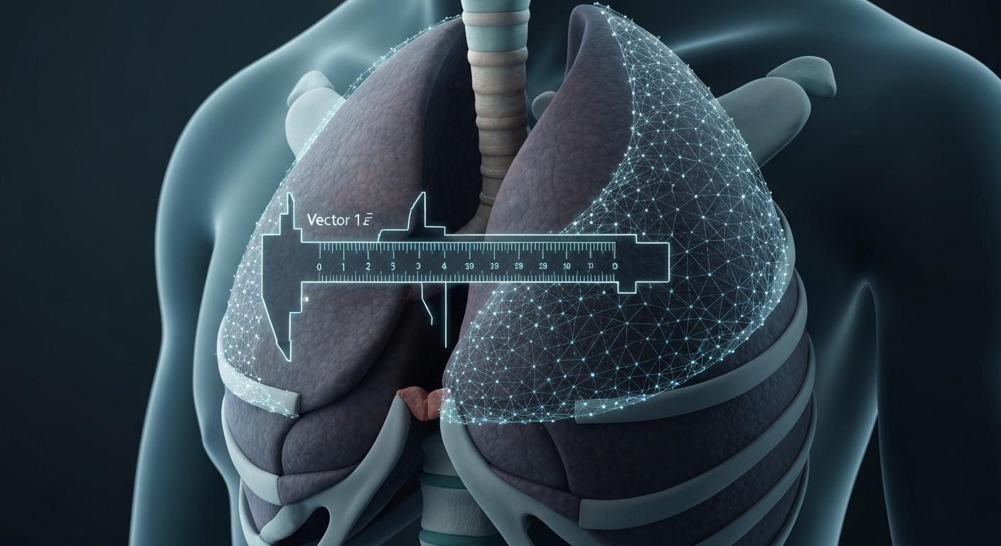
For Doctors in a Hurry
- Clinicians frequently struggle to determine if patients with spontaneous pneumothorax require conservative management or immediate tube thoracostomy.
- The researchers conducted a retrospective study of 101 adult patients to evaluate a vision-enabled artificial intelligence model.
- The model achieved 90.1% accuracy and an area under the curve of 0.90 for predicting necessary clinical interventions.
- The authors concluded that the model provides apical depth estimates and management recommendations consistent with expert radiologist assessments.
- This tool may serve as a clinical decision support system to assist emergency physicians in managing pneumothorax cases.
Quantifying Spontaneous Pneumothorax in the Emergency Department
Managing spontaneous pneumothorax in the emergency department requires rapid differentiation between patients who require immediate tube thoracostomy and those suitable for conservative observation [1]. While digital twins (digital replicas of patient physiology used for real-time monitoring) and advanced sensing technologies are being explored to improve diagnostic precision, clinicians currently rely heavily on manual interpretation of imaging [2, 3]. Deep learning techniques, particularly convolutional neural networks (a class of artificial intelligence designed to process pixel data), have established high accuracy in identifying pulmonary pathologies on chest radiographs, often enhanced by transfer learning (a method where models pre-trained on large datasets are adapted for specific medical tasks) [4, 5]. However, the utility of large language models in radiology remains variable, as some models are prone to fabrication while others demonstrate high reliability for clinical literature synthesis and image segmentation [6, 7]. A new study now evaluates a vision-enabled generative model designed to provide quantitative measurements and specific management recommendations for patients presenting with non-tension spontaneous pneumothorax.
Evaluating Generative Models for Triage Support
The researchers conducted a single-center retrospective observational study to evaluate a vision-enabled generative pre-trained transformer (GPT) model for its ability to estimate apical spontaneous pneumothorax depth and predict initial clinical management. The study population consisted of 101 adult patients (aged 18 years or older) with confirmed spontaneous pneumothorax on posteroanterior chest radiographs. This cohort, identified between January 1, 2023, and December 31, 2025, had a mean age of 33.1 plus or minus 14.6 years and was 88.1% male (n = 89). Among these patients, 53 (52.5%) underwent tube thoracostomy within 24 hours of presentation, providing a real-world baseline for clinical decision-making. To assess the model's utility in an emergency setting, the GPT model received only the chest radiograph image, patient age, and patient sex as inputs. Based on these data, the model produced three specific outputs: the laterality of the pneumothorax, an estimated apical depth in centimeters, and a binary management recommendation of either tube thoracostomy or conservative care. The researchers used the actual performance of a tube thoracostomy within 24 hours of presentation as the reference outcome to validate the model's triage suggestions. This design allowed for a direct comparison between the artificial intelligence's recommendations and the standard of care delivered by emergency department clinicians, bridging the gap between automated image analysis and actionable bedside decisions.
Cohort Characteristics and Clinical Outcomes
The study cohort consisted of 101 patients with a mean age of 33.1 ± 14.6 years, representing a typical adult population presenting to the emergency department with acute respiratory symptoms. Demographic analysis showed that 89 of the 101 patients (88.1%) were male, a distribution consistent with the established higher incidence of spontaneous pneumothorax in men. These baseline characteristics provided the foundation for testing the generative model's ability to process patient-specific data alongside radiographic imaging. To provide a clinical benchmark for the model's triage recommendations, the researchers documented the actual management decisions made by the treating physicians. In the study population, 53 patients (52.5%) underwent tube thoracostomy within 24 hours of their initial posteroanterior chest radiograph. This nearly even split between invasive intervention and conservative management allowed for a rigorous evaluation of the model's predictive accuracy in a high-stakes emergency setting where rapid decision-making is essential for patient safety and resource allocation. Understanding these outcomes is vital for clinicians, as it reflects the real-world complexity of deciding when a lung collapse warrants invasive drainage versus expectant management.
Accuracy in Management Recommendations
The clinical utility of the generative model depends on its ability to distinguish between patients requiring invasive intervention and those suitable for observation. In this cohort, the GPT management recommendations achieved a sensitivity of 86.8% (95% CI, 75.2% to 93.5%), indicating a high rate of correctly identifying patients who required a tube thoracostomy. The model demonstrated even greater performance in identifying patients who did not require intervention, reaching a specificity of 93.8% (95% CI, 83.2% to 97.9%). This high specificity is particularly relevant for emergency department triage, as it suggests the tool could help clinicians avoid unnecessary invasive procedures in stable patients. The overall accuracy for GPT management recommendations was 90.1% (95% CI, 82.7% to 94.5%), reflecting a strong alignment with the final clinical decisions made by the treating physicians. To further validate these findings, the researchers calculated the Cohen's κ (a statistical measure of inter-rater agreement that accounts for the possibility of agreement occurring by chance), which reached 0.80. This value indicates substantial agreement between the model's output and the actual management provided in the emergency department. Additionally, the area under the receiver operating characteristic curve (a metric representing the model's overall ability to discriminate between the need for tube thoracostomy versus conservative care) was 0.90 (95% CI, 0.84 to 0.96). These data points suggest that the vision-enabled GPT can process radiographic and demographic data to produce triage recommendations that closely mirror the standard of care in a high-acuity setting.
Precision in Anatomical Measurement
Spontaneous pneumothorax requires rapid clinical decisions, and the severity of the condition is often dictated by the degree of lung collapse. While artificial intelligence has been applied to thoracic imaging previously, most existing tools focus on the binary detection of a pneumothorax rather than providing the quantitative size estimation necessary for triage. To address this gap, the researchers evaluated the ability of a vision-enabled generative model to measure apical depth, a key metric in determining the clinical pathway. The study compared the model's depth estimates against measurements from blinded radiologists using several statistical benchmarks, including the intraclass correlation coefficient (a descriptive statistic used to assess the consistency of quantitative measurements made by different observers), Bland-Altman analysis, and mean absolute error. The agreement for apical depth estimates between the GPT model and the radiologists was strong, evidenced by an intraclass correlation coefficient of 0.893. This high level of correlation suggests that the generative model can replicate the measurement precision of a specialist when evaluating the extent of a lung collapse on a chest radiograph. Further quantification of the model's accuracy revealed a mean absolute error (the average magnitude of the errors in a set of predictions) of 0.69 cm. In the Bland-Altman analysis (a method used to describe the agreement between two quantitative measurements by plotting the difference between the two against their mean), the mean bias for GPT depth estimation was -0.51 cm. The 95% limits of agreement ranged from -2.30 to 1.27 cm, indicating the range within which most of the differences between the model and the radiologist fall. For the practicing clinician, these metrics provide a clear picture of the model's reliability as an adjunct tool for imaging decision support, offering a standardized method to quantify spontaneous pneumothorax size and assist in the selection of appropriate interventions.
References
1. Günsoy E, Aykut A, Yıldırım C, Öncül MV, Türkoğlu S. Chest X-Ray evaluation using GPT for tube thoracostomy or conservative care in non-tension spontaneous pneumothorax.. Scandinavian journal of trauma, resuscitation and emergency medicine. 2026. doi:10.1186/s13049-026-01616-2
2. Zhang K, Zhou H, Baptista‐Hon DT, et al. Concepts and applications of digital twins in healthcare and medicine. Patterns. 2024. doi:10.1016/j.patter.2024.101028
3. Buttar HM, Rahman MMU, Nawaz MW, Mian AN, Zahid A, Abbasi Q. Non-contact lung disease classification via orthogonal frequency division multiplexing-based passive 6G integrated sensing and communication. Communications Medicine. 2026. doi:10.1038/s43856-025-01181-2
4. Kumar S, Kumar H, Kumar G, Singh SP, Bijalwan A, Diwakar M. A methodical exploration of imaging modalities from dataset to detection through machine learning paradigms in prominent lung disease diagnosis: a review. BMC Medical Imaging. 2024. doi:10.1186/s12880-024-01192-w
5. Ashayeri H, Sobhi N, Pławiak P, Pedrammehr S, Alizadehsani R, Jafarizadeh A. Transfer Learning in Cancer Genetics, Mutation Detection, Gene Expression Analysis, and Syndrome Recognition. Cancers. 2024. doi:10.3390/cancers16112138
6. Güneş YC, Cesur T, Çamur E. Evaluating the reference accuracy of large language models in radiology: a comparative study across subspecialties. Diagnostic and Interventional Radiology. 2025. doi:10.4274/dir.2025.253101
7. Muthukrishnan V, Jaipurkar S, Damodaran N. Continuum topological derivative - A novel application tool for segmentation of CT and MRI images. Neuroimage Reports. 2024. doi:10.1016/j.ynirp.2024.100215