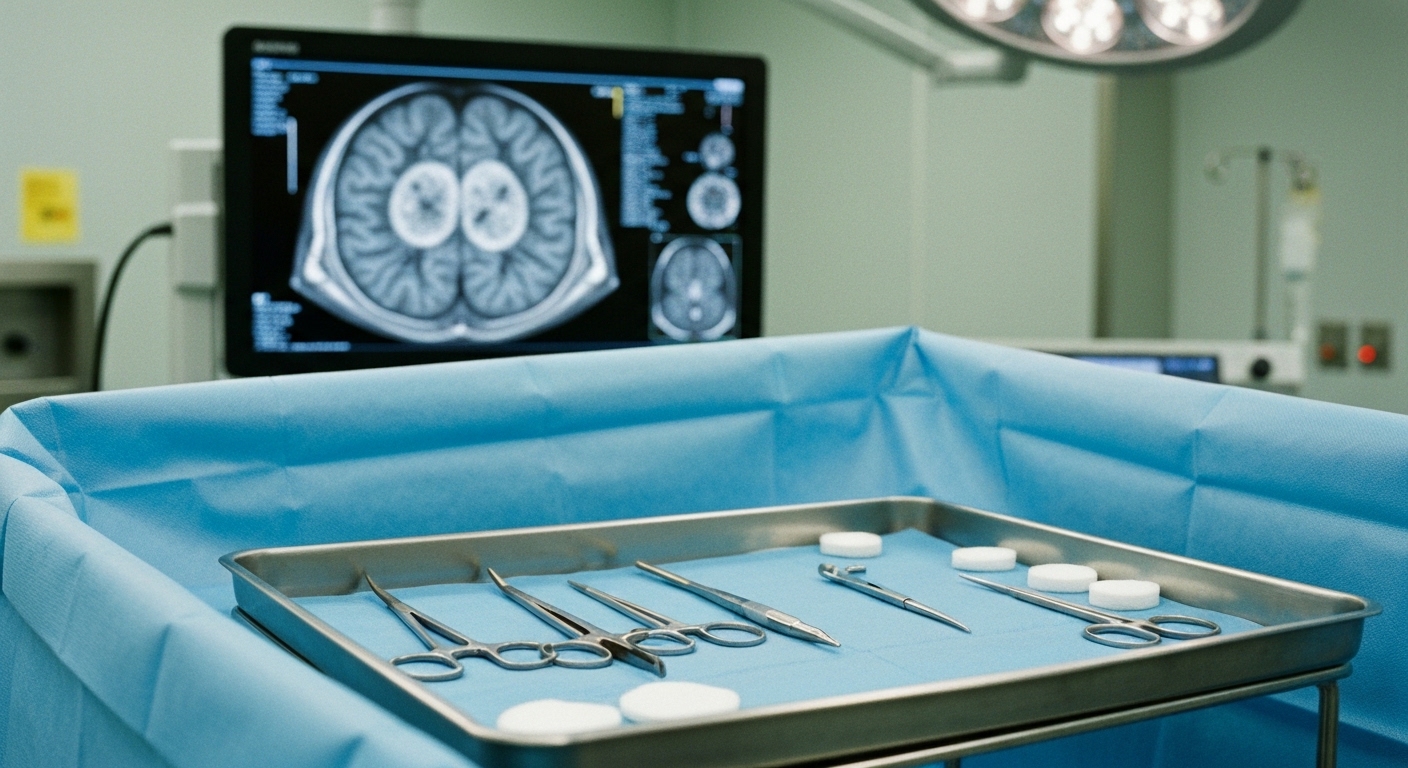
For Doctors in a Hurry
- Current grading systems for brain arteriovenous malformations often fail to account for the specific complexity of eloquent brain regions.
- The researchers retrospectively analyzed 274 patients who underwent microsurgical resection of arteriovenous malformations in critical brain function areas between 2000 and 2025.
- Visual and brainstem locations significantly increased deficit risk, with odds ratios of 14.8 and 19.8 respectively, alongside embedded nidus margins.
- The authors concluded that both anatomical subtype and the degree of margin involvement are critical determinants of postoperative neurological outcomes.
- Clinicians may consider microsurgical resection for unruptured malformations when patients present with preoperative focal neurological deficits, as 27% showed postoperative improvement.
The management of unruptured brain arteriovenous malformations remains a subject of clinical debate, particularly following randomized trials that suggested conservative management might be superior to intervention for certain patients [1, 2, 3]. While microsurgical resection is often utilized to achieve complete lesion removal, its application in areas of critical brain function requires balancing the natural history of hemorrhage against the risk of surgical morbidity [4, 5, 6]. Current grading systems rely heavily on the presence of eloquence (brain regions where injury results in disabling neurological deficits, such as the motor cortex or speech centers), but these models often treat eloquence as a binary variable and may fail to account for anatomical nuances that differentiate one critical region from another [5, 7]. Although multimodal approaches are common, surgery remains a primary method for immediate lesion removal compared to endovascular techniques like curative embolization, which involves the use of liquid embolic agents to obstruct blood flow [8, 9, 6]. A new study now offers fresh insights into how specific anatomical relationships between the lesion and critical brain tissue influence postoperative outcomes.
Defining Critical Brain Function and Nidus Architecture
The researchers conducted a 25-year retrospective cohort study utilizing a prospectively maintained institutional database to evaluate patients treated between 2000 and 2025. The study population consisted of 274 arteriovenous malformations located specifically within areas of critical brain function that underwent microsurgical resection. Among these cases, 171 malformations were ruptured and 103 were unruptured at the time of intervention. By analyzing this longitudinal dataset, the authors sought to move beyond traditional binary grading systems to determine how specific anatomical subtypes and the physical relationship between the lesion and functional tissue influence long term neurological outcomes. To ensure objective classification, the researchers defined the critical brain function subtype based on precise anatomic localization using preoperative T1-weighted and T2-weighted magnetic resonance imaging (MRI). A central focus of the analysis was the degree of involvement between the nidus (the central mass of abnormal vessels where shunting occurs) and the surrounding critical brain tissue.
Quantifying Risk Factors for Postoperative Deficits
The study categorized the relationship between the nidus and functional tissue into two distinct architectural patterns. A nidus was classified as adjacent if any single point of the nidus wall was in contact with a critical brain function region. In contrast, the nidus was defined as embedded if 180 degrees or more of the nidus wall was in contact with critical brain function regions in the axial, sagittal, or coronal planes. This granular definition allows clinicians to better quantify the degree of surgical plane integration and the subsequent risk to functional pathways during resection. The researchers established the primary outcome as the presence of a focal neurological deficit at 2 years postoperatively, providing a long term assessment of functional recovery beyond the immediate surgical window. To identify which variables most accurately predicted these outcomes, the authors utilized a binary logistic regression model (a statistical framework used to predict the probability of a binary outcome based on one or more independent variables). This model proved highly significant (P < 0.001, R2 0.44), demonstrating that both the specific subtype of critical brain function and the degree of margin involvement are primary drivers of postoperative outcomes in patients who were neurologically intact prior to surgery.
Surgical Intervention for Preexisting Neurological Deficits
The analysis identified several independent risk factors that significantly increased the likelihood of new postoperative deficits. The visual critical brain function subtype was associated with a high risk of morbidity (odds ratio 14.8, 95% CI 2.4 to 89.8, P = .002), while the brainstem subtype carried the highest risk among all anatomical locations (odds ratio 19.8, 95% CI 2.0 to 194.6, P = .01). Beyond anatomical location, the physical characteristics of the lesion also played a critical role: nidus size was confirmed as a significant risk factor for new deficits (P = .01). Furthermore, the architectural relationship between the lesion and functional tissue was a major predictor, as an embedded margin significantly increased the risk of a new focal neurological deficit (odds ratio 2.8, 95% CI 1.1 to 7.7, P = .04). The study's analysis of surgical outcomes included a specific subset of 131 patients who were preoperatively intact, meaning they exhibited no focal neurological deficits prior to the procedure. This group was composed of 59 patients with ruptured malformations and 72 patients with unruptured malformations. Beyond those who were neurologically intact, the researchers identified 30 patients with unruptured malformations who presented with preoperative focal neurological deficits, including 21 patients with minor deficits and 9 patients with major deficits. Following microsurgical resection, 8 out of 30 patients (27%) with preoperative deficits from unruptured malformations showed clinical improvement by 2 years postoperatively. This finding is particularly relevant for clinical decision making, as it suggests that the presence of a preoperative focal neurological deficit in an unruptured arteriovenous malformation may be an indication for microsurgical resection rather than a deterrent.
References
1. Luther E, Govindarajan V, McCarthy DJ, et al. Brain Arteriovenous Malformations: Status of Open Surgery after A Randomized Trial of Unruptured Brain Arteriovenous Malformations.. Neurosurgery clinics of North America. 2022. doi:10.1016/j.nec.2022.05.006
2. Volovici V, Schouten J, Gruber A, Meling T, Dammers R. Letter: Medical Management With Interventional Therapy Versus Medical Management Alone for Unruptured Brain Arteriovenous Malformations (ARUBA): Final Follow-up of a Multicentre, Nonblinded, Randomised Controlled Trial.. Neurosurgery. 2020. doi:10.1093/neuros/nyaa448
3. Dmytriw AA, Ku J, Ghozy S, et al. Management of Adult Unruptured Brain Arteriovenous Malformations: An Updated Network Meta-Analysis.. Neurointervention. 2023. doi:10.5469/neuroint.2023.00171
4. Lawton MT. The Role of AVM Microsurgery in the Aftermath of A Randomized Trial of Unruptured Brain Arteriovenous Malformations. American Journal of Neuroradiology. 2014. doi:10.3174/ajnr.a4193
5. Grüter BE, Sun W, Fierstra J, Regli L, Germans MR. Systematic review of brain arteriovenous malformation grading systems evaluating microsurgical treatment recommendation.. Neurosurgical review. 2021. doi:10.1007/s10143-020-01464-3
6. Wong J, Slomovic A, Ibrahim G, Radovanovic I, Tymianski M. Microsurgery for ARUBA Trial (A Randomized Trial of Unruptured Brain Arteriovenous Malformation)-Eligible Unruptured Brain Arteriovenous Malformations.. Stroke. 2017. doi:10.1161/STROKEAHA.116.014660
7. Stuart MJ, Steinberg GK. Prognostic Nuances in the Microsurgical Resection of Arteriovenous Malformations in Areas of Critical Brain Function: A 25-Year Retrospective Cohort Study.. Neurosurgery. 2026. doi:10.1227/neu.0000000000004066
8. Dabhi N, Sokolowski J, Zanaty M, Kellogg RT, Park MS, Mastorakos P. Primary Embolization of Cerebral Arteriovenous Malformations With Intention to Cure: A Systematic Review of Literature and Meta-Analysis. Neurosurgery. 2024. doi:10.1227/neu.0000000000003001
9. Link TW, Winston G, Schwarz JT, et al. Treatment of Unruptured Brain Arteriovenous Malformations: A Single-Center Experience of 86 Patients and a Critique of the A Randomized Trial of Unruptured Brain Arteriovenous Malformations (ARUBA) Trial.. World neurosurgery. 2018. doi:10.1016/j.wneu.2018.09.025