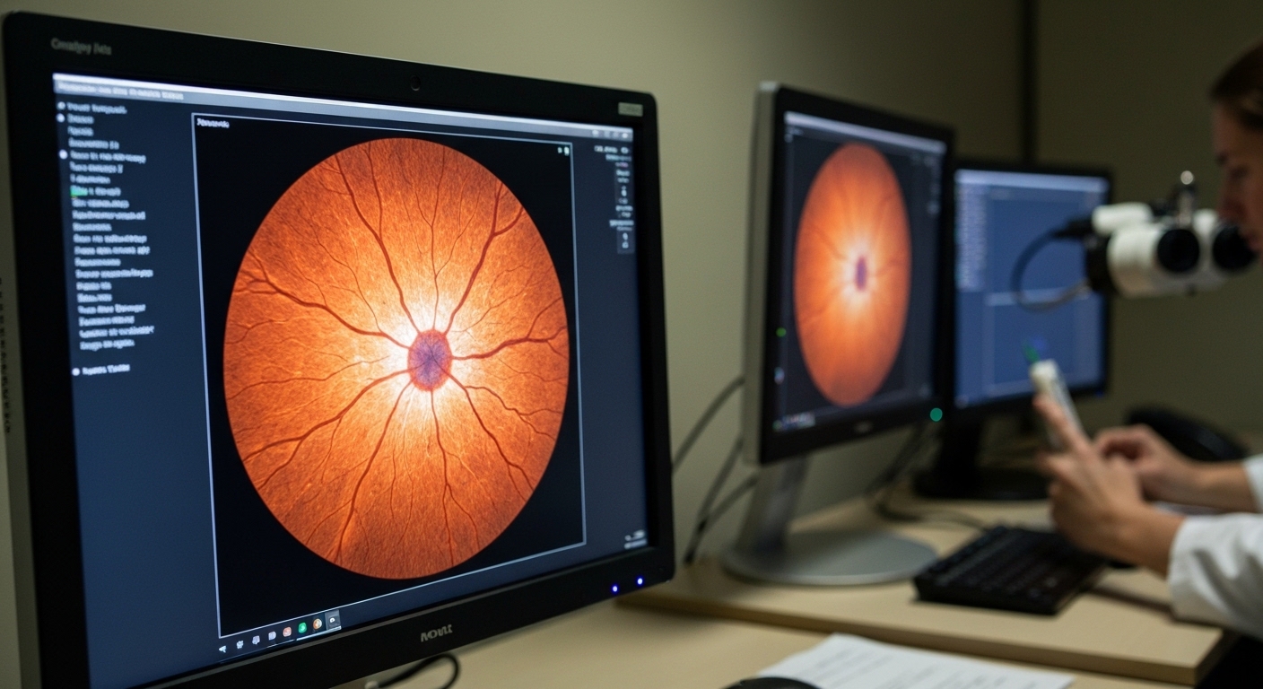
For Doctors in a Hurry
- Clinicians require effective alternatives to panretinal laser photocoagulation for managing proliferative diabetic retinopathy to preserve patient vision.
- This phase 3 trial randomized 689 participants to receive either 6 mg of brolucizumab or standard laser therapy.
- Brolucizumab showed superior visual acuity outcomes with a mean letter score difference of 4.4 at week 54.
- The researchers concluded that brolucizumab effectively preserves visual acuity and reduces retinopathy severity compared to laser treatment.
- Physicians should note that 5.2 percent of brolucizumab patients experienced intraocular inflammation, necessitating careful monitoring during treatment.
Evolution of Management for Proliferative Diabetic Retinopathy
Proliferative diabetic retinopathy remains a leading cause of legal blindness among working-age adults, characterized by pathological neovascularization (the growth of abnormal new blood vessels) that can lead to vitreous hemorrhage and tractional retinal detachment [1]. For decades, panretinal photocoagulation (a laser treatment that ablates peripheral retinal tissue to reduce oxygen demand) has served as the definitive therapy, though it often results in permanent visual field constriction [2, 3]. While intravitreal inhibitors of vascular endothelial growth factor (a protein that stimulates blood vessel growth) have emerged as potent alternatives that can regress neovascularization without tissue destruction, the necessity for frequent injections remains a significant clinical hurdle [4, 5]. Furthermore, the introduction of high-potency agents has raised specific safety concerns regarding intraocular inflammation and retinal vasculitis (inflammation of the retinal blood vessels) [6, 7]. A new study now offers fresh insights into the comparative efficacy and safety of a specific anti-vascular endothelial growth factor dosing regimen versus traditional laser therapy.
Trial Design and Dosing Protocols
The CONDOR trial was a 96-week, 2-arm, single-masked, multicenter, phase 3 randomized clinical trial designed to evaluate the efficacy and safety of brolucizumab, 6 mg, against the standard of care, panretinal photocoagulation (PRP). Conducted across 152 sites in 16 countries, the study analyzed data collected between December 4, 2020, and October 30, 2023, representing a robust international effort to refine the management of advanced diabetic eye disease. The researchers enrolled a total of 689 participants who were randomized in a 1:1 ratio to receive either brolucizumab (n = 347) or PRP (n = 342). Eligible patients were required to have a diagnosis of diabetes and proliferative diabetic retinopathy (PDR), a condition where abnormal blood vessels grow on the retinal surface, and must not have received any previous PRP treatment in the study eye. This treatment-naive population allowed for a direct comparison of pharmacotherapy versus laser without the confounding effects of prior retinal scarring.
The treatment protocol for the brolucizumab arm utilized a specific 6-week loading and 12-week maintenance regimen. Patients received three loading doses of brolucizumab, 6 mg, administered every 6 weeks, followed by maintenance injections every 12 weeks. To address the clinical need for individualized treatment, the protocol allowed for interval extension starting at week 48. Based on disease activity, clinicians could extend the treatment interval by 6 weeks at a time, reaching a maximum of 24 weeks between injections. This flexible approach reflects the real-world clinical goal of reducing treatment burden while maintaining disease control. In contrast, the control group received PRP, which was administered in one to four sessions up to week 12. Following this initial period, patients in the PRP arm could receive additional laser treatment as needed to manage the progression of proliferative disease, mirroring the standard reactive approach to neovascularization.
Baseline Characteristics and Primary Visual Outcomes
The study population consisted of patients with a mean (SD) age of 53.9 (11.39) years, and 276 patients (40.1%) were female. At the start of the trial, the mean (SD) best-corrected visual acuity (BCVA) letter score was 77.1 (10.55), which represents a Snellen equivalent of 20/32. This baseline indicates that participants entered the trial with relatively high functional vision despite their diagnosis of proliferative diabetic retinopathy, making the preservation of that vision a critical clinical priority. Of the total participants randomized at the start of the study, 572 completed the week 54 visit, which served as the primary time point for evaluating visual outcomes.
The primary outcome measure was the change from baseline in best-corrected visual acuity (BCVA) at week 54. The researchers found that brolucizumab was noninferior and superior to panretinal photocoagulation (PRP) for BCVA change at week 54. Specifically, the least-squares mean (SE) change in BCVA letter score was 0.2 (0.72) for the brolucizumab group compared to -4.2 (0.73) for the PRP group. This resulted in a statistically significant difference in BCVA change of 4.4 letters (95% CI, 2.4-6.4; P < .001). These data indicate that while the laser group experienced a loss of nearly one full line of vision on a standard eye chart, the brolucizumab group successfully maintained their baseline acuity. For the practicing clinician, these findings suggest that brolucizumab provides a functional advantage over the traditional standard of care. While panretinal photocoagulation is effective at regressing neovascularization, it is often associated with a gradual decline in visual acuity; in contrast, the 6 mg brolucizumab regimen appears to stabilize vision, offering a viable alternative for patients who prioritize the maintenance of their current visual letter score.
Anatomic Regression and Secondary Endpoints
Beyond the primary visual acuity endpoint, the CONDOR trial evaluated several secondary outcomes, including diabetic retinopathy progression, the prevention of center-involved diabetic macular edema (swelling in the central part of the retina responsible for sharp vision), changes in Diabetic Retinopathy Severity Score levels, and safety. These metrics provide a broader clinical picture of how brolucizumab influences the underlying disease process compared to the thermal destruction of peripheral retina used in panretinal photocoagulation. For the clinician, the prevention of center-involved diabetic macular edema is particularly relevant, as this complication is a frequent cause of vision loss that often requires additional intervention even after successful laser treatment. By targeting the vascular endothelial growth factor pathway, brolucizumab addresses both the proliferative and exudative components of the disease simultaneously.
The anatomic results at one year demonstrated a significant difference in the regression of neovascularization (the growth of abnormal new blood vessels) between the two treatment arms. The researchers found that 187 patients (63.6%) receiving brolucizumab had no proliferative diabetic retinopathy at week 54, whereas only 65 patients (22.4%) receiving panretinal photocoagulation achieved the same status. This represents a difference in the absence of proliferative disease of 39.4% (95% CI, 32.0-46.8; P < .001). These data suggest that the 6 mg brolucizumab regimen is more effective at achieving complete clinical regression of the proliferative signs of the disease than the current standard of care. For practicing ophthalmologists, this high rate of disease regression without the permanent peripheral retinal damage associated with laser therapy may influence the choice of first-line therapy for patients with active neovascularization, particularly those who require high-contrast sensitivity or peripheral vision for their daily activities.
Safety Profile and Inflammatory Risks
The safety profile of brolucizumab in the CONDOR trial provides a critical counterpoint to its visual and anatomic efficacy, offering clinicians a clear view of the trade-offs involved in choosing pharmacotherapy over traditional laser. The researchers reported that ocular adverse events occurred in 119 patients (34.3%) in the brolucizumab arm, whereas a higher proportion of 168 patients (49.1%) in the panretinal photocoagulation arm experienced ocular complications. This difference suggests that the overall burden of ocular side effects may be lower with the 6 mg brolucizumab regimen than with the tissue-destructive nature of laser therapy, which is often associated with permanent peripheral field loss and decreased night vision. However, the specific nature of the adverse events differs significantly between the two modalities.
The study identified specific inflammatory risks that require close clinical monitoring. The rate of intraocular inflammation (an immune response within the eye), including retinal vasculitis (inflammation of the retinal blood vessels), was 18 of 347 patients (5.2%) in the brolucizumab arm. By comparison, the rate of intraocular inflammation was 2 of 342 patients (0.6%) in the panretinal photocoagulation arm. For the practicing physician, these findings establish brolucizumab as a viable alternative to laser for preserving vision, but one that necessitates a higher degree of vigilance for inflammatory complications that are rarely seen with traditional thermal treatment. Clinicians must weigh the superior visual outcomes and higher rates of neovascular regression against the 5.2% risk of intraocular inflammation when counseling patients on their treatment options for proliferative diabetic retinopathy.
References
1. Omar A, Williams G, Whelan J, et al. Diabetic Disease of the Eye in Canada: Consensus Statements from a Retina Specialist Working Group. Ophthalmology and Therapy. 2024. doi:10.1007/s40123-024-00923-0
2. Simmonds M, Llewellyn A, Walker R, et al. Anti-VEGF drugs compared with laser photocoagulation for the treatment of proliferative diabetic retinopathy: a systematic review and individual participant data meta-analysis. Health Technology Assessment. 2024. doi:10.3310/mjyp6578
3. Simmonds M, Llewellyn A, Walker R, et al. Anti-VEGF drugs compared with laser photocoagulation for the treatment of diabetic retinopathy: a systematic review and meta-analysis. Health Technology Assessment. 2024. doi:10.3310/pcgv5709
4. Xie X, Lian C, Zhang Z, et al. Aflibercept for long-term treatment of diabetic macular edema and proliferative diabetic retinopathy: a meta-analysis. Frontiers in Endocrinology. 2023. doi:10.3389/fendo.2023.1144422
5. Virgili G, Curran K, Lucenteforte E, Pető T, Parravano M. Anti-vascular endothelial growth factor for diabetic macular oedema: a network meta-analysis. Cochrane Database of Systematic Reviews. 2023. doi:10.1002/14651858.cd007419.pub7
6. Serhan HA, Taha MJJ, Abuawwad MT, et al. Safety and Efficacy of Brolucizumab in the Treatment of Diabetic Macular Edema and Diabetic Retinopathy: A Systematic Review and Meta-Analysis. Seminars in Ophthalmology. 2023. doi:10.1080/08820538.2023.2271095
7. Lees JS, Dobbin SJ, Elyan B, et al. A systematic review and meta-analysis of the effect of intravitreal VEGF inhibitors on cardiorenal outcomes. Nephrology Dialysis Transplantation. 2022. doi:10.1093/ndt/gfac305