- Researchers investigated if microglial activation and complement-mediated synaptic loss drive the development of cognitive impairment following major surgical procedures.
- This study utilized adult mice undergoing tibial fracture surgery to analyze hippocampal synaptic density and memory performance three days postoperatively.
- Postoperative mice showed a two-fold increase in microglial synaptic protein colocalization (p < 0.001), correlating with significant memory deficits.
- The researchers concluded that hippocampal microglia prune synapses through a C1q-dependent mechanism, directly causing postoperative neurocognitive dysfunction.
- Targeting complement C1q or nuclear factor kappa B activation might prevent the synaptic loss associated with perioperative neurocognitive disorders.
The Pathophysiological Burden of Postoperative Cognitive Decline
Perioperative neurocognitive disorders represent the most frequent neurological complication in geriatric surgical populations, significantly increasing the risk of long-term disability and mortality [1, 2]. Despite affecting millions of patients annually, the underlying pathophysiology remains poorly defined, and clinicians currently lack standardized diagnostic biomarkers or targeted treatments [3, 4]. Current evidence suggests that surgical trauma triggers a cascade of neuroinflammation and mitochondrial dysfunction that disrupts synaptic plasticity [5, 6]. While transient inflammatory insults are known to contribute to these cognitive deficits, the specific mechanisms by which immune cells interact with neuronal architecture postoperatively are still being elucidated [7]. A recent study now offers fresh insights into the molecular signaling pathways that govern these interactions, revealing how the innate immune system actively dismantles brain connections after surgical stress.
Quantifying Cognitive Impairment and Synaptic Loss Post-Surgery
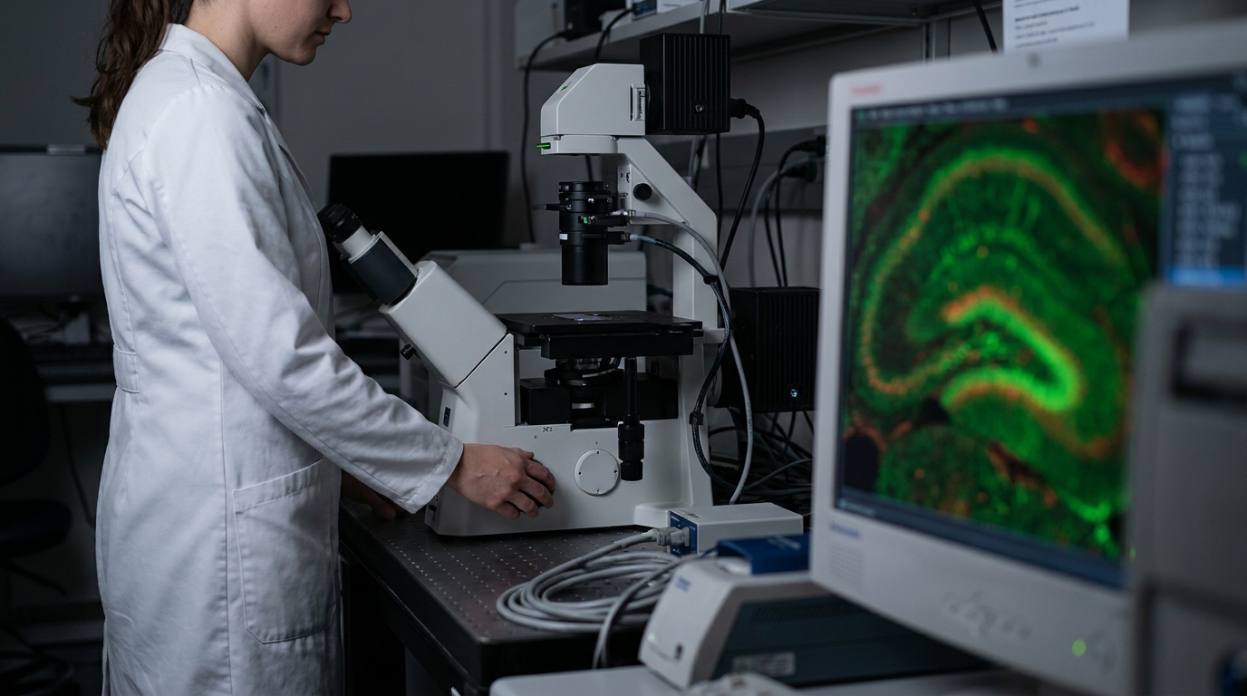
To investigate the cellular drivers of postoperative cognitive decline, researchers utilized a cohort of male and female C57BL/6J mice and male Cx3cr1-CreERT2 mice, all aged between 8 and 12 weeks. These animals underwent aseptic tibial fracture surgery, a procedure designed to simulate the systemic inflammatory response and subsequent neurological impact of major orthopedic surgery in humans. The study employed a comprehensive methodological framework to evaluate hippocampal changes. These included bulk RNA sequencing (a technique used to analyze total gene expression within a tissue sample) to assess global transcriptomic shifts, Western immunoblotting to quantify specific protein levels, and immunofluorescence to visualize cellular interactions. Additionally, the researchers used Golgi staining of the hippocampus, a specialized silver staining method that allows for the high-resolution visualization of individual neuronal dendrites and spines, to assess structural integrity at the synaptic level. Behavioral assessments conducted 3 days after the surgical procedure revealed significant functional impairments that mirror clinical observations of acute confusion and memory loss in elderly patients. The postoperative mice demonstrated marked memory deficits in the Y-maze paradigm (P<0.001), a test evaluating spatial working memory, as well as reduced freezing behavior in the trace fear conditioning paradigm (P=0.002), a sensitive indicator of impaired associative learning. These cognitive declines were closely linked to measurable pathological changes in the brain architecture. Specifically, the researchers observed increased microglial activation (P<0.05) and significant hippocampal synapse loss (P<0.05). For practicing physicians, these findings suggest that the cognitive symptoms observed in the immediate postoperative period are not merely functional disturbances but are rooted in the physical degradation of synaptic networks, likely driven by an overactive immune response within the hippocampal formation.
The Role of Complement C1q in Microglial Phagocytosis
The researchers identified that the observed memory deficits were closely associated with the transcriptomic upregulation of the classical complement pathway (P<0.05), a cascade of proteins typically involved in innate immune responses that has been increasingly implicated in neurodegenerative processes. This genetic shift was accompanied by a significant increase in complement C1q protein levels (P<0.05) within the hippocampal tissue. These findings suggest that surgical trauma initiates a specific molecular signaling pathway that tags healthy synapses for removal by the brain's resident immune cells. Following the surgical procedure, the study observed a ~2-fold increase in the colocalisation of C1qa with Homer1 (P<0.001), an excitatory synaptic protein, as well as a ~2-fold increase in colocalisation with gephyrin (P<0.001), an inhibitory synaptic protein. The researchers defined colocalisation as the physical overlap of microglial markers and synaptic proteins under microscopic imaging, a phenomenon indicating the active engulfment or pruning of these connections by microglia. These data demonstrate that hippocampal microglia prune both excitatory and inhibitory synapses in a C1q-dependent manner. Clinically, this indicates that postoperative cognitive decline results from a broad, structural disruption of hippocampal circuitry rather than a transient imbalance of a single neurotransmitter system, highlighting the profound impact of peripheral surgery on central nervous system integrity.
Targeting the NF-κB/C1q Axis to Preserve Memory
The researchers identified a specific molecular signaling pathway that links surgical trauma to the subsequent loss of hippocampal synapses. The study found that the activation of NF-κB (nuclear factor kappa-light-chain-enhancer of activated B cells), a protein complex that regulates the immune response to infection and stress, correlated with elevated levels of complement C1q in the surgical models. To confirm this relationship, the authors utilized pyrrolidinedithiocarbamate ammonium (PDTC), a selective inhibitor of NF-κB activation. In mice treated with this compound, the selective inhibition of NF-κB activation with PDTC attenuated the surgery-induced elevation of C1q and improved cognitive function, suggesting that the NF-κB pathway is a primary upstream driver of the complement-mediated synaptic pruning observed after tibial fracture. Further pharmacological and genetic interventions provided evidence that blocking this pathway at different stages can preserve neurological function. The researchers observed that treatment with the microglial activity inhibitor minocycline prevented postoperative memory decline and synapse loss, reinforcing the role of microglial activation in the disease process. Similarly, exposure to the C1q neutralizing antibody JL-1 prevented postoperative memory decline and synapse loss, demonstrating that the C1q protein itself is a necessary mediator of the cognitive impairment. To isolate the anatomical site of this pathology, the study showed that C1q depletion from CA1 hippocampal microglia prevented postoperative memory decline and synapse loss, highlighting the CA1 region (a hippocampal area essential for memory consolidation) as a critical site of C1q-dependent synaptic damage. For the practicing clinician, these findings delineate a clear pathophysiological sequence where surgical stress triggers NF-κB signaling, which in turn upregulates C1q and drives microglial phagocytosis of synapses. The fact that selective inhibition of NF-κB with PDTC improved cognitive function in the surgical models suggests that targeting this axis may offer a viable strategy for perioperative neuroprotection. By focusing on the molecular mechanisms of the innate immune system, specifically the classical complement pathway and its regulators, it may be possible to develop targeted pharmacological interventions that mitigate the risk of perioperative neurocognitive disorders in vulnerable surgical populations.
References
1. Tawfik G, Lu MT, Hoz MDL, Crugnola W, Jin Z, Moller DH. Molecular and Clinical Considerations for Anesthesia in the Aging Brain. International Journal of Molecular Sciences. 2025. doi:10.3390/ijms262110272
2. Feng H, Zhang Z, Lyu W, et al. The Effects of Appropriate Perioperative Exercise on Perioperative Neurocognitive Disorders: a Narrative Review. Molecular Neurobiology. 2023. doi:10.1007/s12035-023-03864-0
3. Wiredu K, Aduse-Poku E, Shaefi S, Gerber SA. Proteomics for the Discovery of Clinical Delirium Biomarkers: A Systematic Review of Major Studies. Anesthesia & Analgesia. 2022. doi:10.1213/ane.0000000000006246
4. Dunne SS, Coffey JC, Konje S, et al. Biomarkers in delirium: A systematic review. Journal of Psychosomatic Research. 2021. doi:10.1016/j.jpsychores.2021.110530
5. Zhang Z, Yang W, Wang L, et al. Unraveling the role and mechanism of mitochondria in postoperative cognitive dysfunction: a narrative review. Journal of Neuroinflammation. 2024. doi:10.1186/s12974-024-03285-3
6. Chen B, Qin G, Xiao J, Deng X, Lin A, Liu H. Transient neuroinflammation following surgery contributes to long-lasting cognitive decline in elderly rats via dysfunction of synaptic NMDA receptor. Journal of Neuroinflammation. 2022. doi:10.1186/s12974-022-02528-5
7. Walker KA, Page LML, Terrando N, Duggan MR, Heneka MT, Bettcher BM. The role of peripheral inflammatory insults in Alzheimer’s disease: a review and research roadmap. Molecular Neurodegeneration. 2023. doi:10.1186/s13024-023-00627-2