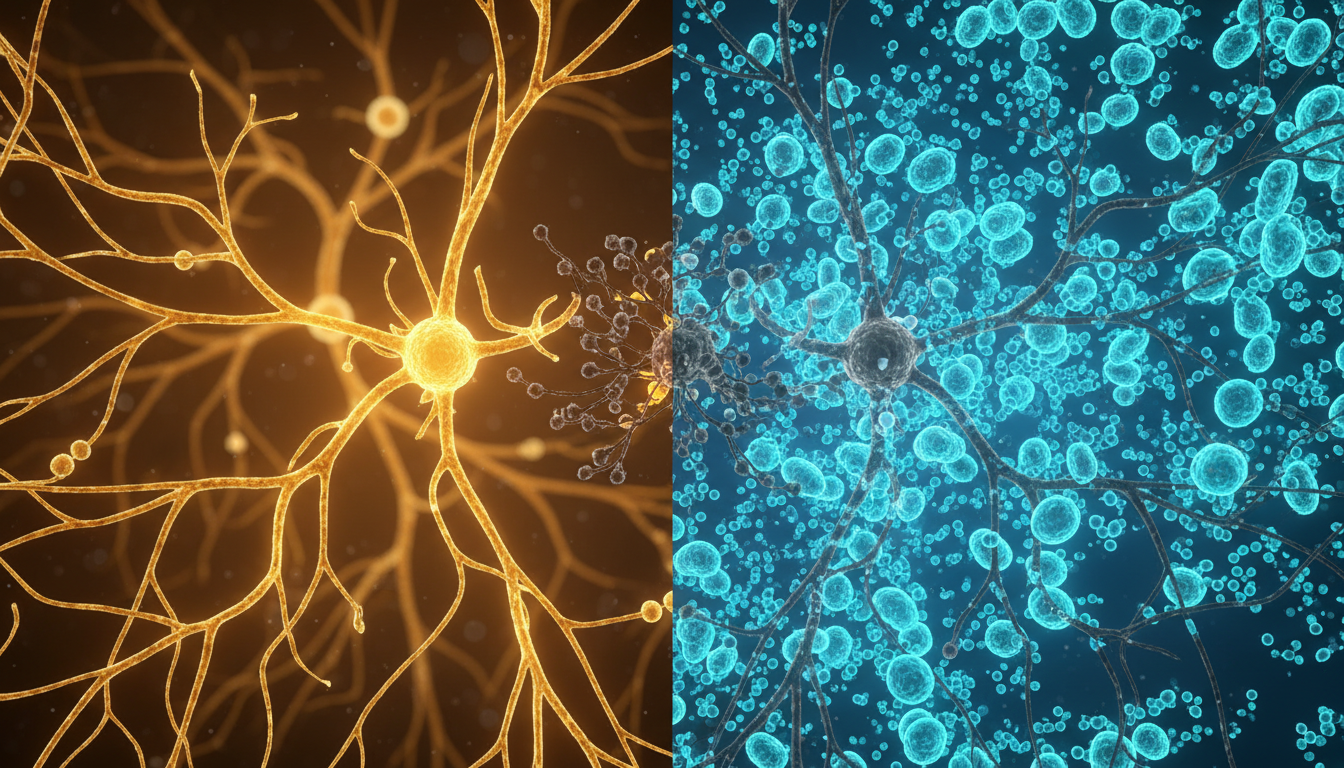
For Doctors in a Hurry
- Researchers investigated whether narcolepsy type 1 is an autoimmune disorder causing the loss of hypocretin, also known as orexin, in the hypothalamus.
- The study examined postmortem brain tissue from patients with narcolepsy type 1 and compared it to control hypothalami to assess immune cell infiltration.
- Postmortem brains demonstrated an 11-fold increase of CD4+ T-cells specifically within the hypocretin region compared with healthy controls.
- The authors concluded that this localized accumulation of CD4+ T-cells expressing memory markers strongly supports the autoimmune hypothesis of narcolepsy type 1.
- Physicians should conceptualize narcolepsy type 1 as a localized autoimmune condition, where targeted CD4+ T-cell activity drives the characteristic loss of hypocretin.
The Elusive Autoimmune Pathology of Narcolepsy Type 1
Narcolepsy type 1 is a chronic neurological sleep disorder characterized by excessive daytime sleepiness and cataplexy, driven by the profound loss of neurons that produce the wake-promoting neuropeptide hypocretin, also known as orexin [1, 2]. For decades, clinicians and researchers have strongly suspected an autoimmune etiology for this neuronal destruction, pointing to the disease's tight association with specific human leukocyte antigen alleles and epidemiological spikes following influenza infections [1, 3]. Despite this compelling circumstantial evidence, directly observing the immune system's assault on the hypothalamus in human patients has proven exceptionally difficult [4, 5]. Consequently, most therapeutic strategies currently rely on managing symptoms rather than addressing the underlying cause of the disease [6]. Now, a recent postmortem analysis of human brain tissue offers a direct look at localized immune activity within the hypothalamus, providing physical evidence that supports the autoimmune hypothesis.
A Highly Localized CD4+ T-Cell Assault
To investigate the cellular mechanisms behind the destruction of hypocretin-producing neurons, researchers analyzed postmortem brains of patients with narcolepsy type 1. The histological examination revealed a highly specific immune infiltration. Specifically, the investigators observed an 11-fold increase of CD4+ T-cells in the hypocretin region of narcolepsy type 1 brains compared with control hypothalami. For clinicians, this massive, localized accumulation of helper T-cells provides a clear physical footprint of the immune system targeting the exact neuronal population responsible for regulating wakefulness. Notably, this immune response was highly selective in its cellular composition. The study found that there was no corresponding rise in CD8+ T-cells in the hypocretin region of affected brains. This absence of cytotoxic CD8+ T-cell proliferation suggests that the pathology is distinctly driven by the CD4+ helper T-cell subset. Furthermore, the researchers discovered that these localized CD4+ T-cells expressed CD49a and CXCR6, which are tissue-resident memory markers (proteins indicating that these immune cells have taken up permanent residence in the brain tissue rather than circulating in the blood). The presence of these specific markers indicates a sustained, localized immune memory within the hypothalamus, reinforcing the autoimmune etiology of the disorder and potentially guiding future targeted immunotherapies.
Sparing the Surrounding Brain Tissue
The spatial specificity of this immune infiltration is striking. While the researchers observed an 11-fold increase of CD4+ T-cells in the hypocretin region of postmortem narcolepsy type 1 brains compared with control hypothalami, without a corresponding rise in CD8+ T-cells, this inflammatory response did not extend to adjacent structures. The study demonstrated that both CD4+ and CD8+ T-cell numbers were unchanged in other hypothalamic regions, including the paraventricular nucleus and median eminence. For clinicians, this indicates that the pathology is not a generalized neuroinflammatory event, but rather a highly targeted assault on a specific neuronal population. This precise targeting extends beyond the immediate vicinity of the hypothalamus. The investigators found that both CD4+ and CD8+ T-cell numbers were unchanged in extra-hypothalamic areas such as the substantia nigra and locus coeruleus. The complete sparing of these critical monoaminergic centers, which are heavily involved in sleep and wakefulness regulation, underscores the extreme selectivity of the neuronal destruction. Ultimately, these findings support the autoimmune hypothesis of narcolepsy type 1, providing concrete physical evidence that the disorder is driven by a localized, antigen-specific immune response rather than a broad neurodegenerative process.
Memory Markers Confirm Autoimmune Etiology
To understand the nature of this localized immune response, the researchers analyzed the molecular phenotype of the infiltrating cells. They discovered that the CD4+ T-cells found in the hypocretin region expressed the tissue-resident memory markers CD49a and CXCR6. For clinicians, the presence of these specific proteins is highly informative. Tissue-resident memory T-cells are a specialized subset of immune cells that, after an initial infection or inflammatory event, take up permanent residence in peripheral tissues rather than circulating in the bloodstream. Their presence indicates a sustained, localized immune memory stationed directly within the brain tissue. This highly restricted spatial distribution of memory T-cells aligns perfectly with the core pathophysiology of the disease. Clinically, narcolepsy type 1 is presumed to be an autoimmune disorder caused by the hypothalamic loss of hypocretin, also known as orexin. The discovery of CD49a and CXCR6-expressing CD4+ T-cells exclusively within the hypocretin fields provides the physical footprint of a chronic, antigen-specific immune assault. Ultimately, these findings support the autoimmune hypothesis of narcolepsy type 1, moving the concept from a strong clinical suspicion to an observable histological reality. By confirming that the destruction of hypocretin-producing neurons is mediated by localized memory T-cells, this research provides a biological rationale for exploring future disease-modifying immunotherapies, rather than relying solely on the symptomatic management of sleep and wakefulness.
References
1. Xu W, Ding W, Zhang Y, et al. The Role of T Cells in the Pathogenesis of Narcolepsy Type 1: A Narrative Review. International Journal of Molecular Sciences. 2024. doi:10.3390/ijms252211914
2. Thorpy MJ, Siegel JM, Dauvilliers Y. REM sleep in narcolepsy. Sleep Medicine Reviews. 2024. doi:10.1016/j.smrv.2024.101976
3. Luo G, Ambati A, Lin L, et al. Autoimmunity to hypocretin and molecular mimicry to flu in type 1 narcolepsy. Proceedings of the National Academy of Sciences. 2018. doi:10.1073/pnas.1818150116
4. Giannoccaro MP, Liguori R, Plazzi G, Pizza F. Reviewing the Clinical Implications of Treating Narcolepsy as an Autoimmune Disorder. Nature and Science of Sleep. 2021. doi:10.2147/nss.s275931
5. Chavda V, Chaurasia B, Umana GE, Tomasi SO, Lu B, Montemurro N. Narcolepsy—A Neuropathological Obscure Sleep Disorder: A Narrative Review of Current Literature. Brain Sciences. 2022. doi:10.3390/brainsci12111473
6. Biscarini F, Barateau L, Pizza F, Plazzi G, Dauvilliers Y. Present and Future of Central Disorders of Hypersomnolence. Journal of Sleep Research. 2025. doi:10.1111/jsr.70118