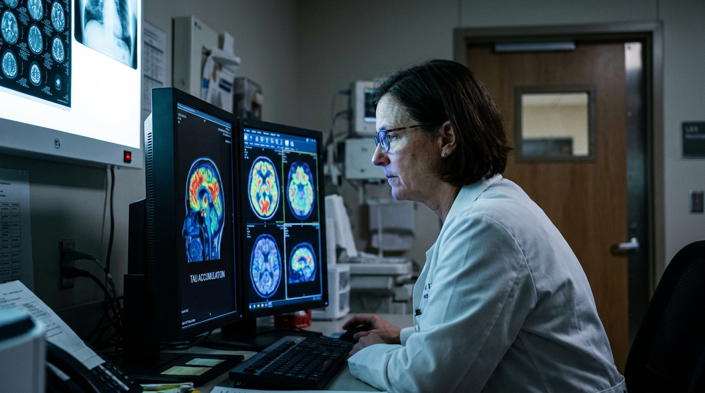
For Doctors in a Hurry
- Researchers investigated whether tau pathology accumulates in non-demented epilepsy patients independently of amyloid pathology or comorbid dementia.
- This study compared 75 epilepsy patients with 47 healthy controls using positron emission tomography imaging and high-throughput plasma proteomics.
- Epilepsy patients showed globally elevated tau tracer uptake across cortical regions despite nonsignificant differences in amyloid-beta levels.
- The researchers concluded that tau accumulation correlates with electroencephalogram abnormalities, clinical disease burden, and accelerated biological aging signatures.
- These findings suggest that tau pathology may serve as a biomarker for disease progression or a target for therapy.
Tau Proteinopathy and the Neurodegenerative Burden of Chronic Epilepsy
Tau protein accumulation is a well-established hallmark of neurodegenerative conditions, traditionally associated with Alzheimer's disease and chronic traumatic encephalopathy [1, 2]. While clinicians have long recognized the cognitive and structural decline that can accompany chronic epilepsy, the underlying molecular drivers of this progression remain poorly defined [3]. Recent evidence suggests that blood-brain barrier dysfunction and reactive astrogliosis, the defense response of astrocytes to central nervous system injury, common in seizure disorders may create a permissive environment for proteinopathy [4, 5, 3]. Furthermore, the interplay between systemic inflammation and microglial activation, the immune response of the brain's resident macrophages, is increasingly viewed as a contributor to accelerated brain aging [6, 7, 8]. To investigate whether these neurodegenerative processes manifest as measurable tau deposition, researchers recently utilized [9]flortaucipir positron emission tomography, a radiopharmaceutical imaging technique that binds to aggregated tau, in a cohort of 75 patients with epilepsy [10].
Widespread Cortical Tau Deposition Independent of Amyloid
The researchers evaluated a cohort of 75 non-demented epilepsy patients and 47 age- and sex-matched healthy controls to investigate the presence of tau pathology. To quantify this burden, they utilized [18F]flortaucipir (FTP) positron emission tomography (PET), a specialized imaging technique that targets and binds to tau protein aggregates in the brain. To ensure the findings were not confounded by typical Alzheimer's disease pathology, a subset of participants also underwent [18F]florbetaben (FBB) PET imaging, which measures the deposition of amyloid-beta plaques. The investigators quantified regional standardized uptake value ratios (SUVRs), a metric of tracer concentration relative to a reference brain region, using the AAL3 brain atlas, a standardized three-dimensional map used to define specific anatomical structures. The imaging results demonstrated that epilepsy patients exhibited globally elevated FTP uptake across multiple cortical regions when compared to the healthy control group. This accumulation of tau was not uniform but was particularly prominent in the lateral and medial frontal, lateral parietal, and lateral occipital brain areas. Notably, the researchers found that FBB SUVRs showed nonsignificant differences between the epilepsy patients and the control group. For practicing neurologists, this lack of significant amyloid-beta deposition suggests that the observed tau accumulation occurs through mechanisms independent of the amyloid-driven pathways typically seen in Alzheimer's disease, pointing toward a distinct pathophysiological process linked directly to chronic epilepsy.
Clinical and Electrophysiological Correlates of Tau Burden
To determine how specific electrophysiological markers correlate with the observed tau burden, the researchers conducted exploratory analyses linking bedside diagnostics to underlying pathology. They found that electroencephalogram (EEG) slowing, a common clinical indicator of cerebral dysfunction or encephalopathy, was associated with higher [18F]flortaucipir (FTP) standardized uptake value ratios (SUVRs). Furthermore, the presence of multifocal discharges on EEG, which represent epileptiform activity originating from multiple independent brain regions, was also associated with higher FTP SUVRs. These findings suggest that the degree of tau accumulation may reflect the severity of underlying neurophysiological disruption and chronic cortical irritability. The study also identified specific clinical history factors that align with increased tau deposition, particularly regarding the timing of seizure activity. Notably, continued seizure activity during adolescence, a critical developmental window for brain maturation and synaptic pruning, was associated with higher FTP SUVRs. This indicates that failing to achieve seizure control during this vulnerable period may exacerbate the long-term accumulation of tau protein. In patients with lateralized epilepsy, where seizures originate from one specific side of the brain, the researchers utilized asymmetry indices, a statistical comparison of tracer uptake between the left and right hemispheres. They found that asymmetry indices for FTP uptake tended to favor the hemisphere with the seizure onset zone. For clinicians, this indicates that tau deposition is not merely a global phenomenon but concentrates in the regions most affected by primary seizure activity, potentially offering a future biomarker for localizing epileptogenic zones.
Proteomic Signatures of Inflammation and Mitochondrial Dysfunction
To investigate the molecular environment associated with tau accumulation, the researchers performed high-throughput plasma proteomic profiling using the SOMAscan platform, a technology that utilizes aptamers to measure thousands of protein levels simultaneously. This analysis identified 473 differentially expressed proteins in patients with epilepsy compared to healthy controls. These proteins were significantly enriched in biological pathways critical to disease progression, specifically those governing immune activation, metabolic regulation, and cytoskeletal remodeling. The identification of these 473 proteins reinforces the concept that chronic epilepsy is not merely a localized electrical disorder but a systemic condition characterized by widespread biochemical alterations. The study further integrated these proteomic findings with neuroimaging data to identify the specific molecular drivers of brain pathology. The researchers measured plasma levels of p-tau217, total tau, and amyloid-beta to complement the PET imaging results. They found that protein expression patterns associated with regional tau standardized uptake value ratios (SUVRs) were specifically linked to immune pathways and mitochondrial dysfunction, the failure of cellular energy production often associated with oxidative stress and neuronal injury. Clinically, these findings suggest that the tau deposition observed on PET scans may be driven by distinct, region-specific mechanisms involving both chronic neuroinflammation and impaired mitochondrial energetics, opening potential avenues for targeted neuroprotective therapies.
Systemic Accelerated Aging and Multi-Organ Impact
The researchers extended their investigation beyond the central nervous system by utilizing the OrganAge algorithm, a statistical tool that estimates biological age based on organ-specific protein signatures found in the plasma. This analysis revealed that epilepsy is associated with accelerated biological aging across several organ systems, most notably in the brain, heart, and muscle. By calculating the brain age gap, which is the difference between an individual's biological age and their chronological age, the study found that epilepsy patients exhibit a physiological profile that is significantly older than their healthy counterparts. These findings suggest that the cumulative burden of chronic epilepsy manifests as a systemic decline in cellular and tissue integrity, rather than an isolated neurological deficit. The study further established a direct link between these systemic aging markers and neurodegenerative pathology. Brain age gaps showed the strongest positive correlation with tau deposition, indicating that patients with the most advanced biological brain aging also harbor the highest levels of tau protein accumulation. This relationship was not confined to the central nervous system; the researchers also found that heart, pancreas, and muscle age gaps correlated with regional brain tau. These correlations suggest that the tau accumulation observed in epilepsy may serve as a broader marker of cumulative biological stress. For the practicing physician, these data imply that the long-term management of epilepsy requires a comprehensive view of the patient's overall health, as the disease correlates with accelerated physiological wear across multiple vital organ systems.
References
1. Hyman BT, Phelps CH, Beach TG, et al. National Institute on Aging–Alzheimer's Association guidelines for the neuropathologic assessment of Alzheimer's disease. Alzheimer s & Dementia. 2012. doi:10.1016/j.jalz.2011.10.007
2. group TT, McKee AC, Cairns NJ, et al. The first NINDS/NIBIB consensus meeting to define neuropathological criteria for the diagnosis of chronic traumatic encephalopathy. Acta Neuropathologica. 2015. doi:10.1007/s00401-015-1515-z
3. Sweeney MD, Zhao Z, Montagne A, Nelson AR, Zloković BV. Blood-Brain Barrier: From Physiology to Disease and Back. Physiological Reviews. 2018. doi:10.1152/physrev.00050.2017
4. Sofroniew MV, Vinters HV. Astrocytes: biology and pathology. Acta Neuropathologica. 2009. doi:10.1007/s00401-009-0619-8
5. Verkhratsky A, Butt AM, Li B, et al. Astrocytes in human central nervous system diseases: a frontier for new therapies. Signal Transduction and Targeted Therapy. 2023. doi:10.1038/s41392-023-01628-9
6. Hoogland IC, Houbolt C, Westerloo DJV, Gool WAV, Beek DVD. Systemic inflammation and microglial activation: systematic review of animal experiments. Journal of Neuroinflammation. 2015. doi:10.1186/s12974-015-0332-6
7. Lawrence JM, Schardien KA, Wigdahl B, Nonnemacher MR. Roles of neuropathology-associated reactive astrocytes: a systematic review. Acta Neuropathologica Communications. 2023. doi:10.1186/s40478-023-01526-9
8. Colonna M, Butovsky O. Microglia Function in the Central Nervous System During Health and Neurodegeneration. Annual Review of Immunology. 2017. doi:10.1146/annurev-immunol-051116-052358
9. Zhang S, Wang X, Gao X, et al. Radiopharmaceuticals and their applications in medicine. Signal Transduction and Targeted Therapy. 2025. doi:10.1038/s41392-024-02041-6
10. Hong SB, Shin Y, Moon J, et al. In vivo tau in epilepsy reflects clinical severity and immune- and ageing-related proteomic changes.. Brain : a journal of neurology. 2026. doi:10.1093/brain/awag145