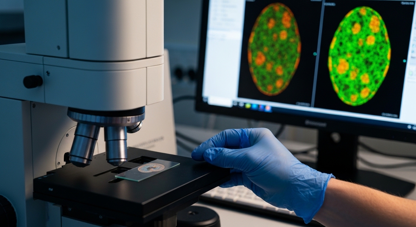
For Doctors in a Hurry
- Researchers investigated the specific brain regions and cellular pathways responsible for sedation induced by the drug dexmedetomidine.
- The study utilized adult male mice to map receptor expression and assess sedation through behavioral and electroencephalography testing.
- Knockdown of astrocyte receptors increased rotarod fall latency to 228 seconds compared to 166 seconds in control subjects (P=0.026).
- The authors concluded that an astrocytic signaling pathway in the hypothalamic paraventricular nucleus mediates the sedative effects of dexmedetomidine.
- These findings suggest that astrocytes are critical targets for future pharmacological strategies aimed at modulating sedation in clinical practice.
The Evolving Neurobiology of Alpha-2 Adrenergic Sedation
Dexmedetomidine has become a cornerstone of modern sedation due to its ability to provide cooperative stability with minimal respiratory depression [1]. It is frequently utilized to reduce the incidence of postoperative delirium in cardiac surgery and to facilitate smoother parental separation in pediatric populations [2, 3]. Despite its clinical utility in intensive care and procedural settings, the drug is associated with dose-dependent bradycardia and hypotension that can complicate hemodynamic management [4, 5]. Current guidelines emphasize its role in opioid-sparing protocols, yet the precise neural circuitry governing its sedative efficacy remains an area of active investigation [6, 7]. A new study now offers fresh insights into the specific cellular pathways that drive these clinical effects, shifting focus from purely neuronal mechanisms to the active role of glial cells.
Mapping Receptor Expression in the Hypothalamus
To clarify the neurobiological mechanisms of dexmedetomidine, a selective alpha-2-adrenergic receptor agonist, researchers conducted a series of experiments using adult male C57BL/6 mice. The study aimed to identify the specific brain regions and cellular pathways that mediate the sedative effects of the drug. By employing RNAscope in situ hybridization (a high-resolution molecular technique used to visualize and quantify specific messenger RNA molecules within intact tissue samples), the team mapped the distribution of alpha-2A-adrenergic receptors across the brain. This technique was combined with immunofluorescence (a method using fluorescently labeled antibodies to detect specific proteins) to pinpoint the exact cellular locations of these receptors. The mapping revealed that alpha-2A-adrenergic receptors were highly enriched in astrocytes within the hypothalamic paraventricular nucleus. While these receptors are traditionally associated with neuronal signaling, the researchers found a dense concentration of the alpha-2A subtype specifically on these non-neuronal glial cells. This finding suggests that the hypothalamic paraventricular nucleus, a region critical for autonomic and endocrine regulation, serves as a central anatomical hub for dexmedetomidine activity, where the drug interacts with astrocytic receptors to initiate the cascade of events leading to clinical sedation.
Quantifying the Loss of Sedative Efficacy
To evaluate the functional impact of astrocytic signaling on dexmedetomidine activity, the researchers utilized three distinct metrics to quantify sedation levels: rotarod performance, open-field locomotion tests, and electroencephalography (EEG). The rotarod test, which measures motor coordination and wakefulness by timing how long a subject can remain on a rotating cylinder, served as a primary behavioral indicator. Complementing this, open-field tests tracked total distance traveled to assess spontaneous locomotor activity, while EEG recordings monitored delta power. In clinical neurophysiology, delta power represents slow-wave brain activity (0.5 to 4 Hz) and serves as a reliable marker of deep sedation and unconsciousness. The study demonstrated that the knockdown of astrocyte alpha-2A-adrenergic receptors significantly reduced the sedative effect of dexmedetomidine, suggesting that these non-neuronal cells are essential for the drug's full clinical potency. Mice with reduced astrocytic receptor expression remained more active and alert following drug administration compared to control groups. Specifically, the knockdown mice showed a significantly increased rotarod fall latency, remaining on the device for a mean (standard deviation) of 228 (52) seconds compared to 166 (62) seconds in the control group (P=0.026). This relative resistance to the drug's effects was further evidenced in the open-field tests, where knockdown mice exhibited an increased travel distance of 3640 (950) cm, whereas controls were limited to 2680 (850) cm (P=0.029). Physiological data from cortical recordings mirrored these behavioral observations, confirming a lighter sedative state at the level of brain circuitry. The EEG delta power decreased in knockdown mice to 54.2 (11.0)% compared to 62.0 (5.9)% in control mice (P=0.024). This reduction in slow-wave activity indicates that the loss of astrocytic alpha-2A-adrenergic receptors prevents the brain from reaching the same depth of pharmacological sedation typically induced by dexmedetomidine. Collectively, these findings establish that the sedative efficacy of this alpha-2-adrenergic agonist is not solely a product of direct neuronal inhibition but is critically dependent on astrocytic modulation within the hypothalamus.
The Role of Astrocytic Calcium and BEST1 Channels
To determine if the alpha-2A-adrenergic receptors identified in the hypothalamus were functional, the researchers utilized intracellular calcium imaging in astrocytes, which are non-neuronal glial cells that support and modulate synaptic activity. This imaging confirmed that dexmedetomidine triggered significant calcium elevations within hypothalamic paraventricular nucleus astrocytes, a process that was strictly dependent on the presence of alpha-2A-adrenergic receptors. This finding suggests that the drug does not merely bypass these cells but actively engages them to initiate a signaling cascade. When the researchers implemented a direct inhibition of astrocyte calcium signaling, they observed a reduction in the sedative response that mirrored the effects seen during receptor knockdown, reinforcing the conclusion that calcium flux within these glial cells is a primary driver of the drug's clinical effect. The study further identified the specific molecular mechanism by which these calcium elevations translate into suppressed neuronal activity. The researchers focused on bestrophin-1 (BEST1), a calcium-activated anion channel that facilitates the release of inhibitory neurotransmitters from astrocytes. By performing a knockdown (a technique that reduces the expression of a specific gene) of bestrophin-1 in the hypothalamic paraventricular nucleus, the team observed a significant impact on the local neurochemical environment. Specifically, the knockdown of bestrophin-1 reduced tonic gamma-aminobutyric acid (GABA) currents to 3.5 (3.4) pA compared to 7.5 (7.2) pA in control subjects (P=0.024). Tonic GABA currents provide a constant level of inhibitory tone to neurons, and their reduction indicates a loss of the inhibitory 'brake' usually applied during sedation. This molecular disruption had direct consequences for the behavioral efficacy of the medication. The knockdown of bestrophin-1 in the hypothalamic paraventricular nucleus significantly diminished the sedative response to dexmedetomidine, as the mice lacked the necessary astrocytic machinery to modulate neuronal firing through GABA release. For the clinician, these data clarify that the sedative properties of dexmedetomidine are mediated by an astrocytic alpha-2A-AR-Calcium-BEST1 signaling pathway. This pathway regulates the inhibitory tone of hypothalamic neurons, suggesting that the depth and quality of pharmacological sedation are as much a product of glial calcium signaling as they are of direct neuronal receptor binding.
Modulating Neuronal Inhibition via GABAergic Tone
The researchers utilized electrophysiology (the study of electrical properties in biological cells and tissues) to examine how astrocytic signaling influences neighboring neurons in the hypothalamic paraventricular nucleus. They specifically targeted Vglut2+ neurons, a population of excitatory cells that utilize glutamate as a neurotransmitter. The study found that dexmedetomidine enhanced tonic gamma-aminobutyric acid (GABA) currents in local Vglut2+ neurons to 15.5 (9.6) pA compared to 7.8 (6.6) pA in control conditions (P=0.035). This increase in tonic GABAergic tone, which provides a constant inhibitory influence on the cell, directly correlated with a reduction in cellular activity. Specifically, dexmedetomidine suppressed neuronal firing in the hypothalamic paraventricular nucleus from a baseline of 1.8 (1.3) Hz down to 0.5 (0.5) Hz (P<0.001). These findings suggest that the sedative effect of the drug is achieved by dampening the activity of excitatory hypothalamic circuits through a sustained inhibitory influence. To confirm the causal link between glial activity and these neuronal changes, the researchers employed chemogenetics (a technique using engineered receptors to precisely control cell activity) alongside astrocyte-specific gene knockdown. They observed that both the enhancement of GABA currents and the suppression of neuronal firing were attenuated when astrocyte calcium levels were inhibited, indicating that the glial calcium surge is a prerequisite for the drug's downstream effects on neurons. This investigation identifies a specific astrocytic alpha-2A-adrenergic receptor-calcium-bestrophin-1 pathway that mediates dexmedetomidine-induced sedation. By characterizing this signaling cascade, the study demonstrates that the clinical state of sedation is not merely a result of direct neuronal receptor binding, but rather a complex interaction where astrocytes modulate the inhibitory environment of the hypothalamus to suppress arousal. For practicing physicians, this highlights the hypothalamic paraventricular nucleus as a key site of action and suggests that future sedative strategies might target glial pathways to achieve desired clinical depths while potentially mitigating side effects.
References
1. Chowdhury S, Sawires J, Weissman B, Saju S, Lambroussis CG. Comparing the Efficacy and Safety of Dexmedetomidine Versus Propofol for Sedation in Adult Patients Undergoing Cardiac Procedures: A Systematic Review.. Cureus. 2025. doi:10.7759/cureus.91773
2. Heybati K, Zhou F, Ali S, et al. Outcomes of dexmedetomidine versus propofol sedation in critically ill adults requiring mechanical ventilation: a systematic review and meta-analysis of randomised controlled trials.. British Journal of Anaesthesia. 2022. doi:10.1016/j.bja.2022.06.020
3. Lang B, Zhang L, Zhang W, Lin Y, Fu Y, Chen S. A comparative evaluation of dexmedetomidine and midazolam in pediatric sedation: A meta-analysis of randomized controlled trials with trial sequential analysis.. CNS neuroscience & therapeutics. 2020. doi:10.1111/cns.13377
4. Gao X, Li Z, Li Z, et al. Is dexmedetomidine superior to non-dexmedetomidine sedatives (particularly propofol) for sedation in critically ill patients with septic shock? A systematic review and meta-analysis of randomized controlled trials. Frontiers in Medicine. 2025. doi:10.3389/fmed.2025.1646256
5. Abosheaishaa H, Abdallfatah A, Abdelghany A, et al. Dexmedetomidine as an Adjunctive Sedative in Patients Undergoing Endoscopic Submucosal Dissection: A Systematic Review and Meta-Analysis. 2025. doi:10.71079/aside.gi.0109257
6. Féray S, Lubach J, Joshi GP, Bonnet F, Velde MVD, Therapy TPWGTESORAAP. PROSPECT guidelines for video‐assisted thoracoscopic surgery: a systematic review and procedure‐specific postoperative pain management recommendations. Anaesthesia*. 2021. doi:10.1111/anae.15609
7. Devlin JW, Skrobik Y, Gélinas C, et al. Clinical Practice Guidelines for the Prevention and Management of Pain, Agitation/Sedation, Delirium, Immobility, and Sleep Disruption in Adult Patients in the ICU. Critical Care Medicine. 2018. doi:10.1097/ccm.0000000000003299