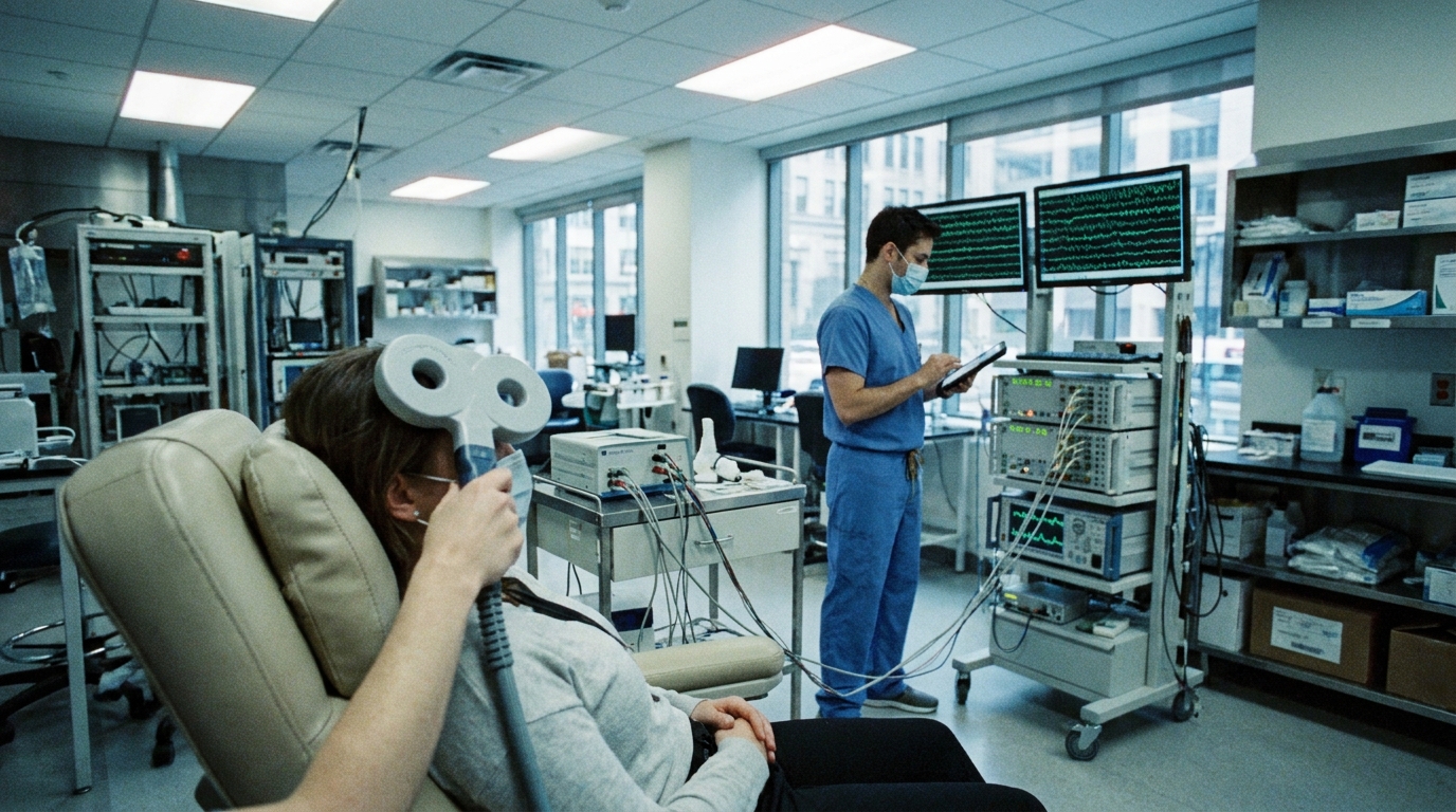
For Doctors in a Hurry
- Researchers investigated how transcranial magnetic stimulation modulates neural circuits by using intracranial electrodes to record direct brain activity.
- This systematic review analyzed seven non-human primate and six human studies utilizing intracranial electroencephalography during magnetic stimulation protocols.
- Single-pulse stimulation induced neuronal excitation within 2 milliseconds, while repetitive protocols produced spectral changes lasting up to 40 minutes.
- The researchers concluded that intracranial recordings effectively map spatial distribution and electrophysiological correlates of stimulation-induced behavioral changes.
- These findings help clinicians understand brain connectivity and functional modulation to refine therapeutic stimulation parameters for neurological disorders.
Refining the Biological Precision of Noninvasive Neuromodulation
Clinicians increasingly utilize repetitive transcranial magnetic stimulation as an adjunctive treatment for drug-resistant epilepsy and major depressive disorder, although the underlying physiological mechanisms remain under investigation [1, 2, 3]. In a meta-analysis of 1,289 patients, low-frequency protocols significantly improved cognitive function (standardized mean difference 1.22; p < 0.0001) and reduced epileptiform discharges (standardized mean difference -0.68; p < 0.00001) [4]. Pediatric data from a randomized controlled trial of 49 children further demonstrate that 0.5 Hz stimulation can achieve a greater than 50 percent seizure reduction in 76 percent of the active group compared to 12.5 percent in the sham group (p < 0.0001) [5]. While these interventions are generally well-tolerated, they carry a crude per-subject seizure risk of 2.9 percent in patients with epilepsy, necessitating a rigorous clinical understanding of how magnetic pulses modulate cortical excitability [6, 7]. Transitioning from broad protocols to targeted therapies requires mapping how external pulses alter internal neural circuits, a process currently being refined through MRI-guided neuronavigation (a tracking system that uses individual imaging to precisely target brain structures) and biomarker-guided strategies [8, 9].
High-Resolution Mapping of Cortical Excitability
To better characterize the physiological impact of neuromodulation, researchers have synthesized data from studies combining transcranial magnetic stimulation (TMS) with direct, invasive brain recordings. Specifically, this review examines the integration of TMS with intracranial micro-stereo electroencephalography (µsEEG) and standard intracranial electroencephalography (iEEG), techniques that utilize electrodes placed directly within or on the surface of the brain. These recording methods provide high spatial and temporal resolution, meaning they can identify the precise anatomical location and the exact millisecond timing of neural activity without the signal interference typically caused by the scalp and skull.
By conducting a systematic search of three major databases, the researchers identified seven non-human primate studies and six human studies that utilized these combined modalities. This cross-species perspective is essential for bridging the gap between animal models and human therapeutic applications, offering direct evidence of how external magnetic fields translate into internal electrical signals within complex neural circuits.
The identified studies employed two primary stimulation formats: single-pulse (spTMS) and repetitive TMS (rTMS) protocols. Single-pulse protocols are typically used to measure the immediate, acute excitability of a specific cortical region, whereas repetitive protocols are designed to induce lasting changes in brain function and connectivity. By analyzing data from both applications, the review clarifies the electrophysiological correlates of brain stimulation, ranging from localized cellular responses to broader shifts in network-wide activity. For the practicing physician, these findings provide a granular map of how therapeutic pulses alter the functional state of the brain in real time, which could eventually guide more precise dosing and targeting in the clinic.
Temporal Dynamics from Milliseconds to Minutes
The exceptional temporal resolution of intracranial recordings allows clinicians to observe the nearly instantaneous cellular response to magnetic stimulation. Data from the reviewed studies indicate that single-unit excitation (the firing of individual neurons) is observed as early as 2 ms after a single TMS pulse. This rapid response confirms that the magnetic field generated by the coil translates into direct electrical activity within the cortical microcircuitry almost immediately upon discharge. By capturing these early events, researchers can map the primary excitatory effects before secondary, polysynaptic signals (activity that has traveled through multiple neuronal connections) begin to influence the recording site.
While single pulses provide insight into acute excitability, repetitive protocols are used to induce more durable physiological changes. The researchers found that intermittent theta-burst stimulation (a specific repetitive TMS protocol involving high-frequency bursts of pulses) can cause neural effects lasting up to 40 min. These sustained changes are characterized by alterations in time-frequency spectra (the patterns of brain wave activity across different oscillation speeds). The fact that these spectral changes persist for 40 minutes after the cessation of stimulation provides a physiological basis for the cumulative therapeutic effects observed in clinical practice.
Understanding this temporal range, from the 2 ms initial excitation to the 40-minute window of spectral modulation, is critical for clinicians optimizing treatment parameters. These findings suggest that the brain remains in a modified state of excitability long after the patient has finished a treatment session. This prolonged window of altered brain wave activity may represent a period of enhanced neuroplasticity, potentially informing the timing of concurrent therapies such as cognitive behavioral interventions or physical rehabilitation. By quantifying these durations, the study offers a more precise framework for determining the necessary frequency and spacing of sessions to maintain therapeutic efficacy.
Local and Remote Network Modulation
The integration of intracranial recordings with magnetic stimulation allows clinicians to visualize the spatial distribution of evoked responses with high precision. The researchers identified that the physiological impact of a pulse is not confined to a single point; rather, the effects include direct influences on neurons near the stimulation site as well as remote effects in regions not directly stimulated. This finding is critical for practicing physicians because it confirms that the therapeutic reach of a focal coil extends through established anatomical pathways to influence distant cortical and subcortical targets. By mapping these connections, the study demonstrates how focal stimulation leads to a broader modulation of brain network interactions, which are the complex communication pathways between different brain areas that govern cognitive and motor functions.
Beyond mapping physical connectivity, these intracranial models enable researchers to investigate the electrophysiological correlates of stimulation-induced modulation, providing the specific electrical signatures that define a brain's response to treatment. This includes the ability to track dynamic changes in network activity as they occur in real time, offering a window into how the brain reorganizes its signaling patterns during and after a session. Most importantly for the clinician, these models can identify the neural correlates that accompany behavioral change, such as improvements in motor speed or mood stabilization. By linking specific electrical patterns to observable clinical outcomes, these findings help bridge the gap between the physics of a magnetic pulse and the biological recovery of the patient.
While these invasive recording techniques provide high-fidelity data, the researchers emphasize that their application requires strict adherence to methodological and safety considerations critical to conducting such studies in both non-human primates and humans. These considerations include the precise placement of micro-stereo EEG and intracranial EEG electrodes to avoid interference with the magnetic field, ensuring that the induced currents do not compromise patient safety or lead to unintended tissue heating. For the practicing clinician, these rigorous protocols underscore the reliability of the data, confirming that the observed changes in neural circuitry are a direct result of the stimulation rather than technical artifacts. Ultimately, this systematic review provides a robust framework for understanding how targeted neuromodulation can be used to predictably alter large-scale brain networks.
References
1. Ong MJY, Rahman MSHA, Chang CJY, et al. Repetitive transcranial magnetic stimulation as adjunctive therapy in adult patients with drug-resistant epilepsy-A systematic review and meta-analysis.. Epileptic disorders : international epilepsy journal with videotape. 2026. doi:10.1002/epd2.70190
2. Lefaucheur J, Alemán A, Baeken C, et al. Evidence-based guidelines on the therapeutic use of repetitive transcranial magnetic stimulation (rTMS): An update (2014–2018). Clinical Neurophysiology. 2020. doi:10.1016/j.clinph.2019.11.002
3. Levkovitz Y, Isserles M, Padberg F, et al. Efficacy and safety of deep transcranial magnetic stimulation for major depression: a prospective multicenter randomized controlled trial. World Psychiatry. 2015. doi:10.1002/wps.20199
4. Xu S, Li S, Yu F, et al. Systematic reviews of low-frequency repetitive transcranial magnetic stimulation on cognition and epileptiform discharge in patients with epilepsy.. PeerJ. 2026. doi:10.7717/peerj.20637
5. Sinha R, Jauhari P, Kumar A, et al. Efficacy of repetitive transcranial magnetic stimulation (rTMS) therapy in children with drug refractory focal epilepsy - A randomized trial.. Seizure. 2025. doi:10.1016/j.seizure.2025.06.020
6. Pereira LS, Müller VT, Gomes MDM, Rotenberg A, Fregni F. Safety of repetitive transcranial magnetic stimulation in patients with epilepsy: A systematic review.. Epilepsy & behavior : E&B. 2016. doi:10.1016/j.yebeh.2016.01.015
7. Allen CH, Kluger BM, Buard I. Safety of Transcranial Magnetic Stimulation in Children: A Systematic Review of the Literature.. Pediatric neurology. 2017. doi:10.1016/j.pediatrneurol.2016.12.009
8. Cooper YA, Pianka ST, Alotaibi NM, et al. Repetitive transcranial magnetic stimulation for the treatment of drug-resistant epilepsy: A systematic review and individual participant data meta-analysis of real-world evidence.. Epilepsia open. 2018. doi:10.1002/epi4.12092
9. Melus J, Waczulikova I, Hapakova L, Konecna M, Valkovic P, Timarova G. Repetitive transcranial magnetic stimulation for the treatment of focal drug-resistant epilepsy: A crossover, randomized, single-blinded, sham-controlled clinical trial.. Seizure. 2025. doi:10.1016/j.seizure.2025.07.015