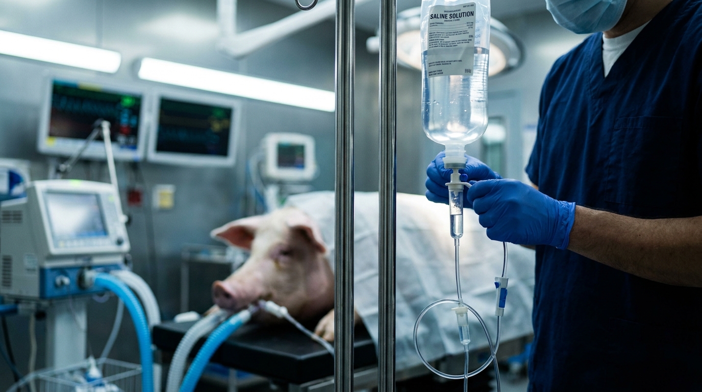
For Doctors in a Hurry
- Researchers investigated whether nicotinamide adenine dinucleotide reduces acute lung injury following cardiac arrest and resuscitation in a porcine model.
- Twenty-two swine were randomized to sham, resuscitation, or resuscitation plus 20 mg/kg intravenous nicotinamide adenine dinucleotide treatment groups.
- Treatment significantly improved the oxygenation index and reduced extravascular lung water compared to the untreated resuscitation group after four hours.
- The study concludes that nicotinamide adenine dinucleotide attenuates pulmonary injury by inhibiting ferroptosis through the signaling of specific regulatory proteins.
- These findings suggest that targeting metabolic pathways may provide a therapeutic strategy for post-resuscitation lung dysfunction in clinical settings.
Mitigating the Pulmonary Toll of Post-Resuscitation Syndrome
The transition from cardiac arrest to successful resuscitation triggers a complex cascade of systemic ischemia-reperfusion injury (tissue damage caused when blood supply returns to organs after a period of oxygen deprivation), often termed post-cardiac arrest syndrome [1, 2]. While return of spontaneous circulation is the primary goal, the subsequent surge in reactive oxygen species (unstable molecules that easily react with other cellular components) and peroxynitrite can cause overwhelming oxidative damage to vital organs [3, 4, 5]. The lungs are particularly vulnerable to this insult, frequently manifesting as acute injury driven by lipid peroxidation and iron-dependent cell death, a process known as ferroptosis [6, 7]. Current clinical management remains largely supportive, as the precise molecular mechanisms governing this pulmonary barrier dysfunction are still being elucidated [5, 2]. A new study in a porcine model investigates whether replenishing metabolic cofactors, specifically nicotinamide adenine dinucleotide (NAD+, a coenzyme central to cellular energy metabolism), can interrupt these lethal signaling pathways to preserve respiratory function after resuscitation [8].
Modeling Post-Arrest Pulmonary Dysfunction
To evaluate the therapeutic potential of nicotinamide adenine dinucleotide (NAD+) in the context of post-resuscitation lung injury, researchers utilized a porcine model involving 22 domestic male swine. The animals were randomly assigned to one of three experimental cohorts: a Sham group (n = 6), a cardiopulmonary resuscitation group (n = 8), and a treatment group receiving both resuscitation and exogenous NAD+ (n = 8). The Sham group underwent surgical preparation only, providing a baseline for physiological comparisons. In the two intervention groups, the researchers established a standardized model of cardiac arrest and cardiopulmonary resuscitation by inducing 10 minutes of ventricular fibrillation followed by 6 minutes of manual chest compressions and ventilations. The clinical intervention was timed to simulate the immediate post-arrest stabilization period. At 5 minutes after the successful return of spontaneous circulation (the restoration of a palpable pulse and blood pressure), the treatment group received intravenous NAD+ at a dosage of 20 mg/kg administered over 1 hour. In contrast, the animals in the Sham and cardiopulmonary resuscitation groups received an equivalent volume of vehicle solution. Hemodynamic stability and survival were monitored closely. Ultimately, six animals in the resuscitation-only group and six animals in the NAD+ treatment group achieved return of spontaneous circulation, allowing for a detailed 24-hour analysis of pulmonary vascular permeability and cellular injury markers.
Preserving Barrier Function and Gas Exchange
To evaluate the integrity of the alveolar-capillary barrier and overall pulmonary function, the researchers utilized several clinical metrics common in intensive care settings. They employed pulse index continuous cardiac output (PICCO) monitoring, a technique that uses transpulmonary thermodilution to provide real-time hemodynamic data, to measure the extravascular lung water index (ELWI) and the pulmonary vascular permeability index (PVPI). The extravascular lung water index serves as a quantitative measure of fluid accumulation within the lung interstitium and alveoli, while the pulmonary vascular permeability index acts as an indicator of how easily fluid leaks from the pulmonary capillaries into the lung tissue. Furthermore, the researchers obtained the oxygenation index (OI) through arterial blood gas analysis to assess the efficiency of gas exchange. To track biochemical evidence of alveolar damage, they determined serum surfactant protein D (SP-D) concentrations, a biomarker that reflects the disruption of the alveolar-capillary membrane, before model induction and throughout a 4-hour post-resuscitation observation period. The data revealed that NAD+ treatment significantly improved lung injury indices, including serum SP-D, ELWI, PVPI, and OI, when compared with the group receiving only cardiopulmonary resuscitation. The stabilization of the alveolar-capillary barrier was evidenced by significant differences in serum SP-D concentrations at multiple time points between the treatment and resuscitation-only groups. The impact on pulmonary edema was particularly pronounced as the post-resuscitation period progressed, with significant differences observed in ELWI and PVPI at the 4-hour mark. For practicing clinicians, these findings indicate that the administration of NAD+ effectively limits the pathological fluid shift and gas exchange impairment that typically complicate the early hours following the return of spontaneous circulation.
Inhibiting Ferroptosis and Cellular Damage
At 24 hours post-resuscitation, the researchers harvested lung tissue samples to conduct a detailed histopathological analysis and an assessment of apoptosis (programmed cell death). These microscopic evaluations revealed that while both the resuscitation-only group and the group receiving NAD+ exhibited marked lung tissue injury and increased apoptosis compared with the Sham group, these pathological changes were significantly attenuated in the group treated with 20 mg/kg of NAD+. Furthermore, the study identified significant differences in lung tissue SP-D levels at the 24-hour mark between the resuscitation-only and treated groups. This finding suggests that the exogenous administration of NAD+ provides a sustained protective effect on the alveolar-capillary unit, reducing the cellular dropout that typically follows a severe ischemic insult. The researchers also investigated the role of ferroptosis, a form of regulated cell death characterized by iron-dependent lipid peroxidation, in post-resuscitation lung injury. Compared with the Sham group, the lung tissue in both intervention groups showed significantly increased levels of ferrous iron (Fe2+), malondialdehyde (MDA, a marker of lipid peroxidation), 4-hydroxynonenal (4-HNE, a toxic byproduct of lipid oxidation), and reactive oxygen species (ROS). Additionally, the expression of acyl-CoA synthetase long-chain family member 4 (ACSL4), an enzyme that promotes ferroptosis by incorporating polyunsaturated fatty acids into cell membranes, was significantly elevated. Conversely, levels of glutathione (GSH), a primary cellular antioxidant, and the expression of Yes-associated protein (YAP), a transcriptional regulator that helps maintain cellular homeostasis, were significantly decreased in both intervention groups compared with the Sham group. Notably, the administration of NAD+ significantly reversed these ferroptosis-related alterations, suggesting that the treatment preserves lung integrity by modulating the YAP/ACSL4 signaling pathway and maintaining the antioxidant capacity of the pulmonary tissue.
The YAP/ACSL4 Signaling Pathway
To elucidate the molecular drivers of pulmonary protection, the researchers evaluated the expression of Yes-associated protein (YAP) and acyl-CoA synthetase long-chain family member 4 (ACSL4) in lung tissue. YAP functions as a transcriptional regulator that promotes cell survival and maintains tissue homeostasis, while ACSL4 is an enzyme that facilitates ferroptosis by catalyzing the esterification of polyunsaturated fatty acids into membrane phospholipids. In the groups subjected to cardiac arrest and resuscitation without treatment, the researchers observed a significant downregulation of YAP and a concomitant upregulation of ACSL4. This imbalance shifts the cellular environment toward a pro-ferroptotic state, contributing to the extensive alveolar damage and capillary leakage observed in the post-resuscitation period. The administration of 20 mg/kg of NAD+ significantly reversed these ferroptosis-related alterations compared with the resuscitation-only group. Specifically, the treatment restored the levels of YAP and glutathione (GSH) while suppressing the accumulation of ferrous iron (Fe2+), malondialdehyde (MDA), 4-hydroxynonenal (4-HNE), and reactive oxygen species (ROS). Furthermore, the treatment effectively inhibited the overexpression of ACSL4. Based on these molecular shifts, the study concludes that NAD+ effectively attenuates lung injury after cardiac arrest and resuscitation in swine by suppressing ferroptosis through the modulation of the YAP/ACSL4 signaling pathway. For the clinician managing post-arrest patients in the intensive care unit, these findings suggest that metabolic interventions targeting specific cell-death pathways could eventually provide a viable strategy to mitigate acute respiratory distress and improve overall survival during the critical early recovery phase.
References
1. Ma X, Liu J, Liu C, et al. ALOX15-launched PUFA-phospholipids peroxidation increases the susceptibility of ferroptosis in ischemia-induced myocardial damage. Signal Transduction and Targeted Therapy. 2022. doi:10.1038/s41392-022-01090-z
2. Wang Y, Hu J, Wu S, et al. Targeting epigenetic and posttranslational modifications regulating ferroptosis for the treatment of diseases. Signal Transduction and Targeted Therapy. 2023. doi:10.1038/s41392-023-01720-0
3. Pacher P, Beckman JS, Liaudet L. Nitric Oxide and Peroxynitrite in Health and Disease. Physiological Reviews. 2007. doi:10.1152/physrev.00029.2006
4. Murphy MP. How mitochondria produce reactive oxygen species. Biochemical Journal. 2008. doi:10.1042/bj20081386
5. Liguori I, Russo G, Curcio F, et al. Oxidative stress, aging, and diseases. Clinical Interventions in Aging. 2018. doi:10.2147/cia.s158513
6. Yan H, Zou T, Tuo Q, et al. Ferroptosis: mechanisms and links with diseases. Signal Transduction and Targeted Therapy. 2021. doi:10.1038/s41392-020-00428-9
7. Ru Q, Li Y, Chen L, Wu Y, Min J, Wang F. Iron homeostasis and ferroptosis in human diseases: mechanisms and therapeutic prospects. Signal Transduction and Targeted Therapy. 2024. doi:10.1038/s41392-024-01969-z
8. Förstermann U, Sessa WC. Nitric oxide synthases: regulation and function. European Heart Journal. 2011. doi:10.1093/eurheartj/ehr304