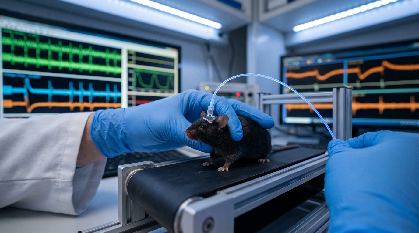
- Researchers investigated how physical activity modulates the balance of excitatory and inhibitory signals within specific hippocampal CA1 neuron populations.
- The study utilized voltage imaging and optogenetics to monitor pyramidal cells and three distinct types of inhibitory interneurons.
- Locomotion reduced firing in pyramidal cells and vasoactive intestinal peptide interneurons while increasing activity in somatostatin and parvalbumin cells.
- The authors concluded that locomotion induces a divisive gain reduction, which scales down neuronal output, through balanced excitatory and inhibitory inputs.
- These findings clarify how behavioral states alter hippocampal processing, which may inform future treatments for movement-related cognitive dysfunction.
Cellular Dynamics of Hippocampal Circuit Integration
The hippocampus serves as a critical hub for memory consolidation and spatial navigation, processes heavily influenced by the internal state and external behaviors of a patient [1, 2]. Clinical understanding of hippocampal function centers on the balance between excitatory and inhibitory signaling, a disruption of which is implicated in epilepsy and neurodevelopmental conditions such as autism spectrum disorder [3, 4, 5]. While traditional electrophysiology has mapped general activity, the specific ways neuronal subtypes integrate signals through subthreshold dynamics (the membrane potential changes occurring before a neuron reaches its firing threshold) remain difficult to isolate in vivo [4]. Recent advances in high-density recording and neuromodulatory tracking, such as the use of genetically encoded dopamine sensors, have begun to clarify how circuits adapt to real-time sensory and motor inputs [6, 7]. A recent study utilizes all-optical electrophysiology (a method using light to simultaneously stimulate neurons and record their electrical activity) to demonstrate that locomotion reduces firing in pyramidal cells and vasoactive intestinal peptide interneurons while increasing activity in somatostatin and parvalbumin cells [8].
Divergent Behavioral Responses Across Interneuron Subtypes
The researchers utilized voltage imaging and optogenetics to monitor the activity of four distinct neuronal populations in the CA1 region of the hippocampus. These included pyramidal cells (PCs), which serve as the primary excitatory neurons; vasoactive intestinal peptide (VIP) interneurons, which typically disinhibit the circuit; somatostatin (SST) interneurons; and parvalbumin (PV) interneurons. The study observed that the transition from rest to locomotion triggered a divergent response among these subtypes. Specifically, locomotion was found to reduce firing in pyramidal cells (PCs) and reduce firing in vasoactive intestinal peptide (VIP) interneurons. This suppression of excitatory output and its primary disinhibitory regulators suggests a significant reorganization of hippocampal processing during physical activity.
In contrast to the suppression observed in pyramidal cells and VIP cells, the researchers found that locomotion increased activity in somatostatin (SST) interneurons and increased activity in parvalbumin (PV) interneurons. From a clinical perspective, this shift is highly relevant because SST and PV cells provide critical inhibitory control over the dendrites and somata (the cell bodies) of pyramidal neurons, respectively. The simultaneous increase in these inhibitory populations during movement likely serves as a regulatory mechanism to prevent runaway excitation, a phenomenon often seen in seizure disorders, and to maintain the precision of spatial encoding. By mapping these firing rate changes, the study illustrates a shifting circuit landscape where the brain selectively suppresses certain signals while enhancing others to modulate the gain (the ratio of output to input) of hippocampal computations during active states.
Inhibitory Contributions to Theta Oscillations
To investigate the cellular mechanisms underlying hippocampal rhythms, the researchers utilized a sophisticated platform that combined voltage imaging with optogenetics to examine genetically defined CA1 neurons in the hippocampus. This approach allowed for the simultaneous recording of membrane potential and the precise manipulation of neuronal activity. A key component of the experimental protocol involved the use of prolonged optical depolarization, a technique where light is used to steadily increase the membrane potential of a neuron to evaluate the contribution of inhibitory inputs to its overall activity. By elevating the baseline voltage of these cells, the authors could more clearly observe how inhibitory signals from neighboring interneurons shaped the fluctuations in the electrical state of the cell.
The study focused specifically on the origins of intracellular theta oscillations, which are rhythmic fluctuations in membrane potential (typically occurring at 4 to 12 Hz) that organize the timing of hippocampal activity and are essential for memory encoding. The findings revealed that inhibitory inputs contribute substantially to intracellular theta oscillations in pyramidal cells (PCs). Furthermore, the researchers determined that these same inhibitory inputs contribute substantially to intracellular theta oscillations in vasoactive intestinal peptide (VIP) cells. For the practicing clinician, these results clarify that theta rhythms are not merely the result of rhythmic excitatory drive but are actively sculpted by inhibitory signaling. This mechanism ensures the temporal precision of neuronal firing, which is necessary for the hippocampus to accurately process spatial and temporal information during behavior and may explain cognitive deficits seen when inhibitory networks degrade in neurodegenerative diseases.
Selective Gain Control and Somatodendritic Balance
To determine how physical activity alters the relationship between input and output in the hippocampus, the researchers analyzed how excitatory (E) and inhibitory (I) inputs modulate spiking output, subthreshold dynamics, and gain. The team utilized firing rate-laser intensity (F-I) curves to reveal distinct gain modulation across cell types, a method that measures the change in the firing rate of a neuron in response to increasing levels of optogenetic stimulation. This analysis allowed the researchers to quantify gain, which is the process by which a neuron scales its output in response to varying input intensities. The data showed that locomotion caused a divisive gain reduction in pyramidal cell (PC) bursting, effectively dampening the high-frequency clusters of action potentials that are often associated with the transmission of high-fidelity information. In contrast, simple spikes in pyramidal cells (PCs) remained unaffected by locomotion-induced gain modulation, suggesting that the brain selectively filters specific types of neural messages during movement.
The researchers employed a two-compartment model to analyze the underlying mechanism of these effects, simulating the distinct electrical environments of the cell body and the branching extensions. The model suggests the gain reduction results from a balanced increase in excitatory/inhibitory (E/I) input to the soma (the cell body) as well as a balanced increase in excitatory/inhibitory (E/I) input to the dendrite. This simultaneous increase in both excitation and inhibition acts as a stabilizing force, allowing for precise control over neuronal output without saturating the activity of the cell. These findings demonstrate how behavior coordinates excitatory/inhibitory (E/I) signaling to modulate hippocampal computations, providing a cellular basis for how the brain prioritizes different streams of information during active navigation. For the clinician, this highlights a sophisticated level of circuit regulation where the E/I balance does not just maintain stability but actively reconfigures how the hippocampus processes spatial and temporal data based on the behavioral state of the patient, offering potential new targets for modulating circuit dysfunction in psychiatric and neurological disorders.
References
1. Rasch B, Born J. About Sleep's Role in Memory. Physiological Reviews. 2013. doi:10.1152/physrev.00032.2012
2. Buzsáki G. Hippocampal sharp wave‐ripple: A cognitive biomarker for episodic memory and planning. Hippocampus. 2015. doi:10.1002/hipo.22488
3. Jiang C, Lin L, Long S, et al. Signalling pathways in autism spectrum disorder: mechanisms and therapeutic implications. Signal Transduction and Targeted Therapy. 2022. doi:10.1038/s41392-022-01081-0
4. Valero M, Zutshi I, Yoon E, Buzsáki G. Probing subthreshold dynamics of hippocampal neurons by pulsed optogenetics. Science. 2022. doi:10.1126/science.abm1891
5. Kuner R, Kuner T. Cellular Circuits in the Brain and Their Modulation in Acute and Chronic Pain. Physiological Reviews. 2020. doi:10.1152/physrev.00040.2019
6. Patriarchi T, Cho JR, Merten K, et al. Ultrafast neuronal imaging of dopamine dynamics with designed genetically encoded sensors. Science. 2018. doi:10.1126/science.aat4422
7. Steinmetz NA, Aydın Ç, Lebedeva A, et al. Neuropixels 2.0: A miniaturized high-density probe for stable, long-term brain recordings. Science. 2021. doi:10.1126/science.abf4588
8. Yang Q, Baror-Sebban S, Kipper R, London M, Adam Y. All-optical electrophysiology reveals behavior-dependent dynamics of excitation and inhibition in the hippocampus. bioRxiv. 2025. doi:10.1101/2025.03.20.644347