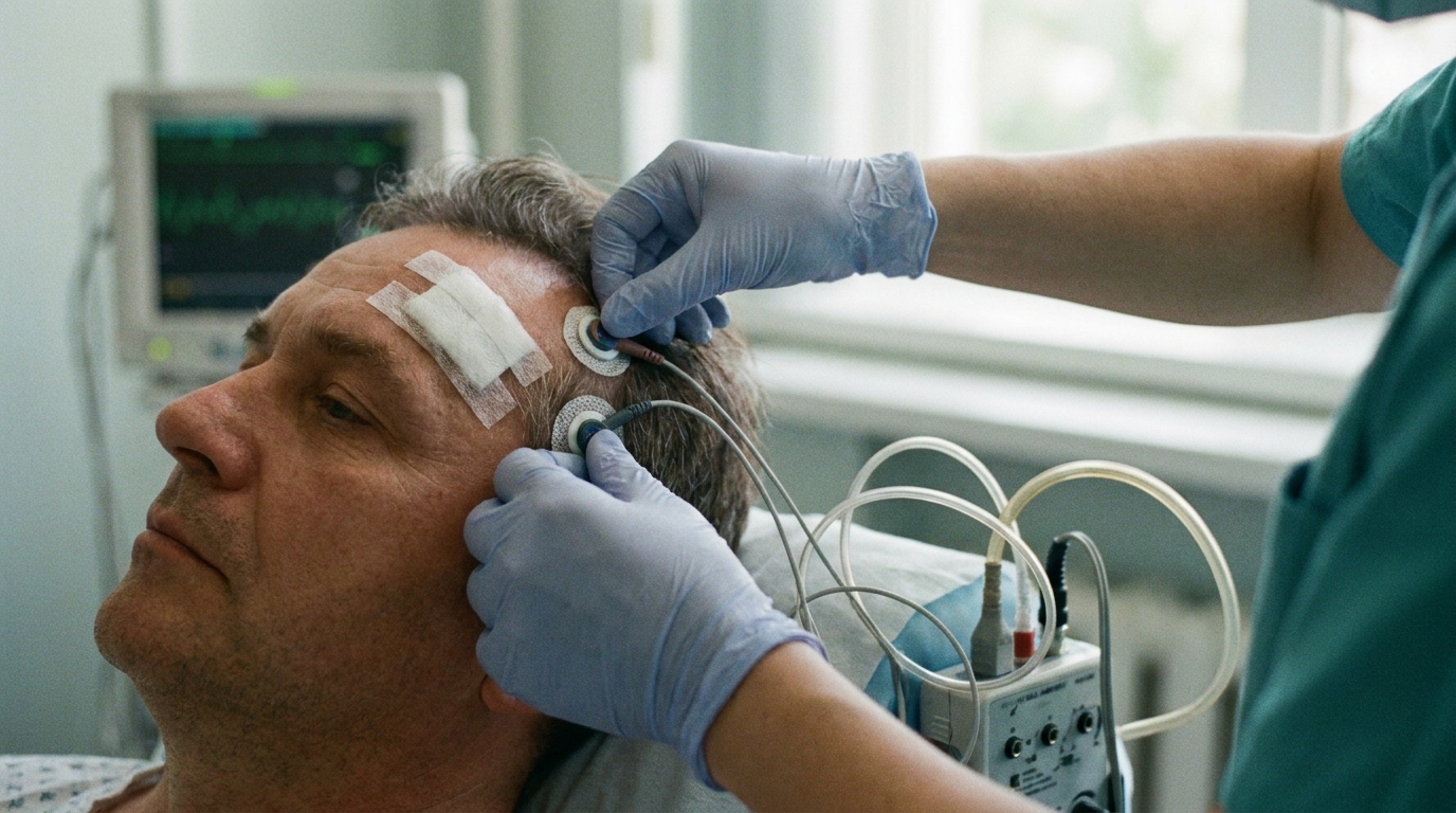
For Doctors in a Hurry
- Researchers investigated whether medial temporal lobe lesions specifically impair the precision of visual working memory or the total quantity of retained information.
- This study evaluated 40 patients with drug-resistant epilepsy, comparing 19 individuals with hippocampal lesions to 21 controls before and after surgery.
- Hippocampal damage significantly increased recall variability, while total lesion volume, not hippocampal extent, predicted a lower recall success rate.
- The researchers concluded that the medial temporal lobe preserves the fidelity of memory representations rather than the overall quantity of stored content.
- Measuring visual working memory precision may provide a sensitive clinical marker for monitoring functional decline in patients with focal brain lesions.
Redefining Memory Deficits in Temporal Lobe Epilepsy
Surgical intervention remains a primary treatment for drug-resistant focal epilepsy, often involving the resection of the hippocampus or surrounding medial temporal structures [1, 2]. While these procedures are effective for achieving seizure freedom, they carry inherent risks of neuropsychological morbidity, particularly regarding memory and cognitive processing [3]. Current diagnostic frameworks emphasize the structural and genetic heterogeneity of these epileptogenic lesions [4, 5]. However, the specific impact of focal medial temporal damage on the nuances of visual working memory (the ability to briefly hold and manipulate visual information) remains a subject of debate in clinical neurology. A recent study of 40 surgical cases demonstrated that lesions specifically involving the hippocampus significantly increased recall variability, indicating that such damage reduces the precision or fidelity of visual representations rather than the total quantity of information retained [6]. This distinction offers clinicians a more precise way to counsel patients on expected postoperative cognitive changes.
Dissociating Memory Quality from Quantity
To investigate the specific cognitive architecture of memory deficits, researchers examined visual working memory recall performance in 40 patients with drug-resistant epilepsy before and after brain surgery. This longitudinal design allowed for a direct comparison of cognitive function following surgical intervention. Within this cohort, the investigators identified 19 patients with lesions involving the hippocampus, a structure traditionally associated with long-term memory consolidation but increasingly implicated in short-term processing. The remaining 21 patients served as a comparative group, having either no lesions or lesions located entirely outside the hippocampus. This division enabled the team to isolate the functional contributions of the medial temporal lobe from the effects of general surgical trauma or broader cortical damage.
The experimental protocol utilized a controlled visual working memory task with a fixed set size (presenting a consistent number of items to remember), designed to minimize non-target recall errors where a patient might accidentally report the features of an incorrect item. Using this framework, the researchers mathematically modeled participants' responses to distinguish between two distinct components of memory. They modeled recall variability as an inverse measure of visual working memory precision, reflecting the clarity or fidelity of the stored information. Simultaneously, they modeled the probability of recall success as the proportion of trials not attributable to failed, uniform recall responses, serving as a metric for memory capacity or the total number of items successfully retained. For practicing physicians, this distinction is vital because it separates a patient's ability to remember that an object existed from their ability to remember its exact characteristics, such as its precise color or orientation.
Hippocampal Damage and Reduced Recall Fidelity
Analysis of the 19 patients who underwent surgical resection involving the medial temporal lobe showed that lesions affecting the hippocampus led to a significant increase in recall variability. This metric is clinically relevant because increased recall variability indicates reduced visual working memory precision after surgery, meaning patients could still remember that an item existed but were less accurate regarding its specific features. Unlike general cognitive decline, this deficit specifically targets the fidelity of the mental representation rather than the total number of items a patient can hold in mind.
To pinpoint the anatomical source of these deficits, the researchers employed voxel-based lesion-symptom mapping (a statistical technique that correlates tissue damage in specific three-dimensional brain coordinates with clinical deficits). This high-resolution analysis revealed a robust association between hippocampal damage and increased recall variability, confirming that the hippocampus plays a specialized role in maintaining the resolution of visual information. Notably, the association between hippocampal damage and recall variability remained significant even after controlling for overall brain lesion volume. This suggests that the loss of precision is a localized consequence of hippocampal injury rather than a byproduct of the total amount of brain tissue removed during surgery.
While broader surgical impact (measured as total lesion volume) predicted the overall rate of recall success, it did not predict the precision of those memories. Only specific damage to the hippocampal structure correlated with increased noise in visual recall. Consequently, neurologists and neuropsychologists evaluating postoperative patients should recognize that hippocampal integrity is specifically required for memory sharpness, not just memory presence.
Global Lesion Burden and Recall Success
To distinguish between the specific functional role of the hippocampus and the impact of more extensive neurological damage, the investigators analyzed the relationship between overall lesion size and memory performance. While hippocampal injury specifically degraded the resolution of memories, the study found that total lesion volume predicted a reduced recall success rate. This suggests that the probability of successfully retrieving any information at all is tied to the cumulative loss of brain tissue rather than the focal destruction of medial temporal structures. In this context, recall success refers to the proportion of trials where a patient could provide a response that was not a random guess, reflecting the basic capacity to hold an item in mind.
The data further clarified that hippocampal lesion extent did not predict the recall success rate, reinforcing the conclusion that the hippocampus is not the primary driver of memory quantity. Instead, the findings indicate that a broader lesion burden constrains how much content is retained, resulting in more failed recall responses. While the hippocampus ensures that a memory is sharp and distinct, the overall integrity of the wider cortical network appears to determine whether the memory is present or absent. For the practicing clinician, this means that a patient with a large surgical resection may struggle to remember that an object was shown to them at all, whereas a patient with a small, targeted hippocampal lesion may remember the object but fail to recall its exact color or orientation.
Clinical Implications for Cognitive Assessment
Historically, classic lesion case-control studies suggested that the medial temporal lobe had minimal involvement in visual working memory, particularly when patients were asked to remember simple stimulus features such as color or orientation. However, more recent intracranial recordings have implicated the medial temporal lobe, and specifically the hippocampus, in supporting visual working memory precision (the resolution or sharpness of a stored representation). The hippocampus supports this precision by distinguishing between similar visual features, a process that reduces representational variability during short retention intervals. Because medial temporal lobe activity scales with visual working memory set size (the number of items a person is asked to remember), some researchers previously suggested the region might contribute to both the quality and the quantity of retained content.
This study specifically addressed models that posit a unitary memory strength metric (a theoretical framework using a single value to represent overall memory performance, accounting for both the quantity and quality of recall). The authors found that while an alternative model assuming this unitary metric could capture the overall performance decline associated with increasing total lesion volume, it could not account for the specific effects observed in the medial temporal lobe. These results challenge existing cognitive models that treat visual working memory quality and quantity as interchangeable consequences of a single underlying memory strength parameter. Instead, the findings highlight that the medial temporal lobe is essential for preserving the fidelity of representations rather than their mere presence.
For clinicians managing patients with focal brain lesions, these findings suggest that standard binary assessments of memory (which only measure whether a patient remembers an item or not) may be insufficient. Because the researchers identified distinct neural correlates for memory quantity and quality, visual working memory precision may serve as a sensitive behavioral marker for tracking functional changes. Incorporating precision-based tasks into neuropsychological evaluations could allow for more nuanced monitoring of cognitive status in individuals with memory impairment, detecting the subtle increase in recall variability that occurs when the hippocampus can no longer effectively distinguish between similar visual inputs.
References
1. Darko MK, Ohenewaa P, Tenkorang B‡O, et al. Epilepsy Surgery for Drug-Resistant Epilepsy in Africa: A Systematic Review. Neurosurgery. 2024. doi:10.1227/neu.0000000000003307
2. Gong M, Xu K, Shan Y, et al. Protocol of a prospective multicenter randomized controlled trial of robot-assisted stereotactic lesioning in the treatment of focal drug-resistant epilepsy. Trials. 2023. doi:10.1186/s13063-023-07334-9
3. Stern MA, Malcolm J, Willie J, Gross R, Drane D. Letter: Magnetic Resonance Imaging-Guided Laser Interstitial Thermal Therapy for Epilepsy: Systematic Review of Technique, Indications, and Outcomes.. Neurosurgery. 2020. doi:10.1093/neuros/nyaa274
4. Blümcke I, Thom M, Aronica E, et al. The clinicopathologic spectrum of focal cortical dysplasias: A consensus classification proposed by an ad hoc Task Force of the ILAE Diagnostic Methods Commission1. Epilepsia. 2010. doi:10.1111/j.1528-1167.2010.02777.x
5. Najm I, Lal D, Alonso‐Vanegas M, et al. The ILAE consensus classification of focal cortical dysplasia: An update proposed by an ad hoc task force of the ILAE diagnostic methods commission. Epilepsia. 2022. doi:10.1111/epi.17301
6. Choo Y, Thakurdesai SP, Qadri A, et al. Medial temporal lobe lesions reduce visual working memory precision.. 2025. doi:10.1093/brain/awaf397