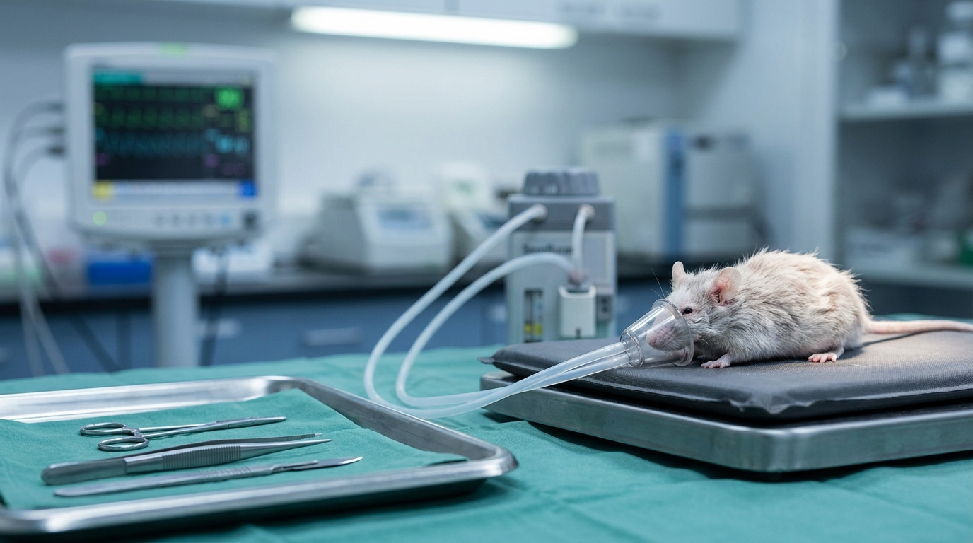
For Doctors in a Hurry
- Researchers investigated how disruptions in hippocampal sharp-wave ripples (electrical patterns for memory consolidation) contribute to postoperative memory impairment in elderly patients.
- This study used 18-month-old mice exposed to sevoflurane and laparotomy to model surgical effects on hippocampal neural activity and memory.
- Inducing sharp-wave ripples improved memory consolidation from 34.3 percent to 53.1 percent (P=0.016) while restoring CA3 pyramidal neuron activity.
- The researchers concluded that surgery-induced microglial activation inhibits CA3 neurons, which disrupts sharp-wave ripple dynamics and impairs memory consolidation.
- These findings suggest that targeting neuroinflammation or hippocampal electrical patterns may mitigate cognitive decline following major surgery in older patients.
Deciphering the Neural Basis of Postoperative Cognitive Decline
Postoperative cognitive dysfunction and delirium remain significant challenges for clinicians managing elderly patients, with incidences reaching 18.4 percent following elective procedures [1]. These complications are associated with increased mortality, longer hospital stays, and a higher likelihood of discharge to skilled nursing facilities [2, 3]. While various pharmacological strategies, such as dexmedetomidine or specific anesthetic choices, have been explored to mitigate these risks, the underlying pathophysiology of memory-specific deficits remains poorly understood [4, 5]. Because current guidelines often lack the mechanistic specificity required to develop targeted prevention strategies for surgery-induced cognitive decline [6], researchers recently investigated the exact hippocampal circuit disruptions that occur following surgical stress.
Surgical Stress and Memory Consolidation Deficits
To investigate the mechanisms underlying postoperative cognitive decline, researchers utilized 18-month-old male C57BL/6J mice, an animal model selected to represent the physiological state of elderly patients. These mice were subjected to a standardized surgical protocol consisting of 3 vol% sevoflurane anesthesia combined with a laparotomy to simulate the systemic stress of a major abdominal procedure. This experimental design allowed the authors to isolate the effects of surgical trauma and anesthesia on the aging brain, focusing specifically on how these stressors impact the hippocampal circuits responsible for cognitive function. The study identified that postoperative memory impairment in these aged mice was specifically associated with deficits in memory consolidation, the physiological process by which unstable, newly acquired information is converted into long-term storage. This failure to stabilize memories was linked to distinct electrophysiological changes in the hippocampus, particularly within the CA1 region (an area critical for memory formation). The researchers observed a reduced frequency and duration of sharp-wave ripples (SPW-Rs), which are high-frequency electrical oscillations essential for the stabilization and transfer of new memories. By quantifying these events, the authors demonstrated that the disruption of these specific hippocampal signals serves as a primary physiological marker for the observed cognitive deficits following surgery.
Restoring Memory Through Ripple Induction
To investigate the physiological mechanisms underlying these cognitive deficits, the researchers employed a multi-modal experimental framework. They utilized context fear conditioning (a behavioral task where animals learn to associate a specific environment with an aversive stimulus) to assess memory retention. This was paired with local field potential monitoring, a technique involving implanted electrodes to record the collective electrical activity of neuronal populations, allowing for the real-time tracking of hippocampal oscillations. Furthermore, the team used immunofluorescence staining to visualize specific cellular changes and protein expression within the hippocampal architecture. The study specifically focused on whether the observed reduction in SPW-Rs was a causal factor in postoperative cognitive decline or merely a secondary symptom. By using optogenetic and chemogenetic tools (techniques that use light or engineered drugs to precisely control genetically modified neurons) to artificially trigger these high-frequency oscillations, the researchers tested if they could bypass the surgical disruption. The data demonstrated that the induction of SPW-Rs improved memory consolidation scores from 34.3 (9.4) percent to 53.1 (18.7) percent (P=0.016). These findings establish a direct link between the frequency of hippocampal ripples and the ability of the brain to stabilize new information following anesthesia and surgery. For the clinician, this highlights a specific electrophysiological target for understanding postoperative delirium and memory loss. The results indicate that the cognitive impairment observed in the aged mice is driven by a reversible disruption in hippocampal signaling, providing a physiological basis for future interventions aimed at preserving cognitive function in elderly surgical patients.
The CA3 to CA1 Circuit Failure
To identify the upstream mechanisms driving this hippocampal dysfunction, the researchers employed viral tracing, a technique using modified viruses to map anatomical connections between neurons. This allowed for a precise investigation of the signaling between the CA3 and CA1 hippocampal regions. The study found that surgical stress led to a significant decrease in the number of c-Fos-positive pyramidal neurons in the CA3 region, where the c-Fos protein serves as a cellular marker for recent neuronal activation. This reduction in CA3 pyramidal neuron activity directly contributed to diminished excitatory transmission to the CA1 region, effectively starving the CA1 of the input necessary to generate robust SPW-Rs. The researchers then tested whether restoring activity in this circuit could reverse the observed deficits. By using chemogenetic approaches to selectively activate CA3 pyramidal neurons, they successfully restored the activity of downstream CA1 pyramidal neurons. This intervention ameliorated the disruption of SPW-R frequency, increasing it from 0.20 (0.05) to 0.25 (0.15) events per second (P=0.018). Furthermore, the duration of these ripples improved from 0.034 (0.0028) to 0.039 (0.0033) seconds (P=0.014). Most importantly for clinical considerations, the targeted activation of CA3 neurons was sufficient to ameliorate postoperative memory impairment in the aged mice. These results suggest that the cognitive decline seen after surgery is mediated by a specific failure in the excitatory drive from the CA3 to the CA1 region, a circuit that remains functionally salvageable through targeted neuromodulation.
Microglial Neuroinflammation as the Primary Driver
The researchers identified microglial activation, which refers to the recruitment and inflammatory response of the resident immune cells in the brain, as a central driver of the observed hippocampal dysfunction. In the 18-month-old mice, the physiological stress of surgery triggered a robust immune response. This microglial activation was associated with SPW-R disruption and postoperative memory deficits, suggesting that the immune system plays a direct role in the failure of neural circuits. The study found that surgery triggers microglial activation, leading to the release of neuroinflammatory factors into the hippocampal environment. These signaling molecules specifically inhibit the activation of hippocampal CA3 pyramidal neurons, the primary excitatory cells responsible for driving signal transmission to the CA1 region. The inhibition of CA3 neurons disrupts SPW-R dynamics and impairs memory consolidation, effectively preventing the brain from stabilizing new information into long-term storage. For the practicing clinician, these findings clarify the pathological pathway from the operating room to cognitive decline: surgical trauma induces a neuroinflammatory state that silences the CA3-CA1 circuit. This mechanism suggests that targeting microglial activation or the resulting inflammatory cascade may be a viable future strategy for preserving hippocampal circuit integrity and preventing postoperative cognitive complications in geriatric patients.
References
1. Watt J, Tricco AC, Talbot‐Hamon C, et al. Identifying Older Adults at Risk of Delirium Following Elective Surgery: A Systematic Review and Meta-Analysis. Journal of General Internal Medicine. 2018. doi:10.1007/s11606-017-4204-x
2. Bulut H, Lam C, Patel RH, et al. Outcomes of modern total joint arthroplasty in patients with dementia: a systematic review of challenges and considerations for perioperative care. Archives of Orthopaedic and Trauma Surgery. 2026. doi:10.1007/s00402-026-06233-3
3. Greaves D, Psaltis PJ, Davis D, et al. Risk Factors for Delirium and Cognitive Decline Following Coronary Artery Bypass Grafting Surgery: A Systematic Review and Meta‐Analysis. Journal of the American Heart Association. 2020. doi:10.1161/jaha.120.017275
4. Zhang H, Lü Y, Liu M, et al. Strategies for prevention of postoperative delirium: a systematic review and meta-analysis of randomized trials. Critical Care. 2013. doi:10.1186/cc12566
5. Meineke MN, Applegate RL, Rasmussen T, et al. Cognitive dysfunction following desflurane versus sevoflurane general anesthesia in elderly patients: a randomized controlled trial. Medical Gas Research. 2014. doi:10.1186/2045-9912-4-6
6. Weidmann AE, Proppé GB, Matthíasdóttir R, Tadić I, Gunnarsson P, Jónsdóttir F. Medication-induced causes of delirium in patients with and without dementia: a systematic review of published neurology guidelines. International Journal of Clinical Pharmacy. 2025. doi:10.1007/s11096-024-01861-4