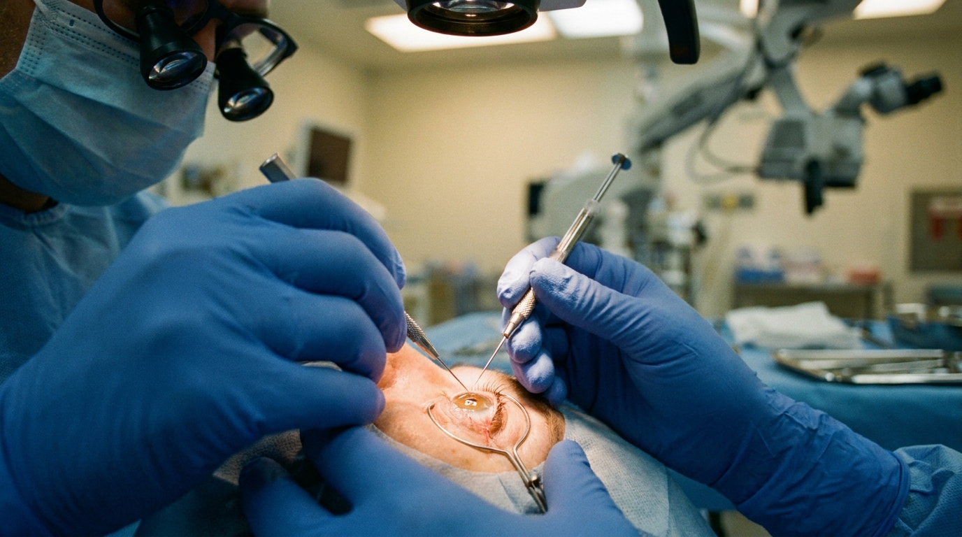
For Doctors in a Hurry
- Traditional surgeries for primary angle-closure glaucoma provide intraocular pressure control but involve significant risks and prolonged recovery periods.
- This review evaluates anatomical and clinical evidence regarding minimally invasive glaucoma surgery as a safer alternative for managing angle-closure.
- Clinical evidence indicates that these procedures achieve 20 to 50 percent reductions in intraocular pressure while decreasing medication dependency.
- The researchers conclude that targeted interventions can complement or replace invasive filtration surgery by restoring physiological outflow through multiple mechanisms.
- Future advancements will focus on imaging-guided precision and device refinements to enable individualized, mechanism-based treatment pathways for patients.
Evolving Surgical Strategies for Primary Angle Closure Glaucoma
Primary angle-closure glaucoma remains a significant clinical challenge due to its high rate of irreversible vision loss and complex anatomical drivers, such as pupillary block and plateau iris configuration (a condition where the iris is pushed forward by the ciliary body, narrowing the drainage angle) [1, 2]. While phacotrabeculectomy (combined cataract extraction and traditional filtration surgery) is the conventional standard for advanced cases, it carries a 23.7% complication rate and requires intensive postoperative management [3, 4]. Minimally invasive glaucoma surgery has emerged as a safer alternative, with meta-analyses of 875 patients demonstrating a weighted mean intraocular pressure reduction of 7.71 mmHg (95% CI: 5.16 to 10.26) and a significant decrease in medication burden [5]. Specific techniques like phacogoniotomy have shown non-inferiority to phacotrabeculectomy in pressure reduction (26.1 mmHg versus 25.7 mmHg) while offering shorter operative times and improved quality of life scores [3]. Other approaches, including gonioscopy-assisted transluminal trabeculotomy (an ab interno procedure that opens the trabecular meshwork to restore physiological drainage), achieve a 76% success rate in angle-closure eyes, providing clinicians with mechanism-based options to refine surgical decision-making and tailor interventions to individual patient anatomy [6].
Anatomical Drivers of Angle Closure
Primary angle-closure glaucoma represents a critical global health concern as a major cause of irreversible blindness worldwide. Epidemiological data indicate that the highest disease burden is concentrated in Asia, where anatomical predispositions to narrow angles are most prevalent. For the practicing clinician, managing this condition requires a nuanced understanding of its multifactorial pathogenesis, which involves the complex interplay of several distinct mechanical drivers rather than a single physiological failure.
The primary mechanisms of angle closure include pupillary block, a condition where the iris adheres to the lens and physically blocks the flow of aqueous humor from the posterior to the anterior chamber. This obstruction is often compounded by plateau iris configuration, in which the iris is pushed forward by the ciliary body, directly narrowing the drainage angle. Additionally, lens-related crowding occurs when the crystalline lens occupies excessive space in the anterior segment, further reducing the available volume for aqueous circulation. Recent evidence also highlights the role of dynamic choroidal expansion, characterized by increased volume in the vascular layer of the eye, which can acutely exacerbate angle narrowing.
Recognizing these specific drivers allows for a shift from generalized surgical protocols to targeted interventions. By identifying whether a patient's condition is primarily driven by pupillary block, plateau iris, lens-related crowding, or choroidal expansion, surgeons can better utilize minimally invasive techniques to address the specific anatomical obstruction. Clinical evidence suggests that combining these targeted approaches with lens extraction can achieve intraocular pressure reductions of 20% to 50%, offering a mechanism-based alternative to more invasive filtration surgeries and allowing physicians to preserve vision with fewer surgical risks.
Limitations of Conventional Filtration Surgery
The historical standard of care for managing primary angle-closure glaucoma has relied on a specific set of invasive interventions. Traditional surgical management includes trabeculectomy, lens extraction, or transscleral cyclophotocoagulation (a procedure utilizing laser energy to treat the ciliary body and reduce the production of aqueous humor). While these techniques are frequently utilized in advanced cases, they are often viewed as a final resort due to the physiological trauma associated with creating a full-thickness drainage fistula or ablating secretory tissue.
Clinical evidence indicates that these traditional surgeries achieve substantial intraocular pressure control, which is vital for halting the progression of a disease that remains a leading cause of irreversible blindness. However, the high efficacy of these procedures is coupled with a demanding clinical profile. These conventional methods carry significant risks of serious complications, including hypotony, endophthalmitis, and bleb-related issues. Furthermore, patients undergoing these procedures often require prolonged recovery periods, necessitating frequent postoperative monitoring and intensive topical regimens that can complicate the management of a busy clinical practice.
Mechanisms and Efficacy of Minimally Invasive Techniques
Minimally invasive glaucoma surgery (MIGS) expands treatment options for primary angle-closure glaucoma once angle access is restored, providing clinicians with safer, earlier, and targeted interventions tailored to the underlying anatomical mechanisms of the disease. Unlike traditional filtration surgeries that create a new drainage pathway, MIGS focuses on enhancing or bypassing existing physiological routes. These techniques are categorized by their anatomical targets, including procedures that address the trabecular meshwork (the primary drainage tissue) and Schlemm's canal (the circular channel that collects aqueous humor). Other approaches involve the placement of subconjunctival stents to redirect fluid or ciliary body approaches that modulate aqueous production at its source.
The clinical utility of these procedures is significantly enhanced when combined with lens extraction. This dual approach addresses the mechanical drivers of angle closure by deepening the anterior chamber and releasing peripheral anterior synechiae, which are pathological adhesions between the iris and the drainage angle. By clearing these obstructions, the combined procedure restores physiological outflow through the natural drainage pathways. Clinical evidence demonstrates that these interventions achieve intraocular pressure reductions of 20% to 50%. Furthermore, the implementation of these techniques allows for a reduced medication burden, enabling patients to maintain target pressures with fewer topical drops while avoiding the high complication rates associated with more invasive filtration surgeries.
Integrating MIGS into Clinical Practice
The clinical utility of minimally invasive glaucoma surgery is shifting the management of primary angle-closure glaucoma toward more individualized, mechanism-based treatment pathways. The researchers suggest that these procedures can complement or, in selected cases, replace invasive filtration surgery such as trabeculectomy. By integrating these techniques earlier in the disease process, clinicians can potentially avoid the high risk profiles and prolonged recovery periods associated with traditional filtration while still achieving significant intraocular pressure control. This transition allows for a more nuanced approach where the surgical intervention is tailored to the specific anatomical drivers of the patient's glaucoma, such as lens-related crowding or trabecular meshwork resistance.
The continued evolution of this field depends on several technical and pharmacological advancements. Future progress is contingent upon device refinements specifically designed for narrow-angle eyes, which present unique spatial challenges for traditional instrumentation. Furthermore, the authors highlight the necessity of imaging-guided precision to ensure accurate device placement and to monitor anatomical changes in the drainage angle in real time. These hardware improvements must be paired with biologic modulation (the use of pharmacological agents to control the healing response and prevent the fibrosis that can lead to surgical failure).
Beyond technical specifications, the widespread adoption of these procedures requires a focus on strategies to enhance cost-effectiveness and accessibility. Because primary angle-closure glaucoma disproportionately affects populations in resource-limited settings, particularly in Asia, reducing the economic burden of these devices is critical for global implementation. By combining these economic strategies with improved clinical outcomes, the researchers aim to establish a sustainable model for glaucoma care that prioritizes early intervention and long-term safety over the more reactive, high-risk approaches of the past.
References
1. Song Y, Song W, Zhang Y, et al. Efficacy and safety of phacotrabeculectomy versus phacogoniotomy in advanced primary angle-closure glaucoma: study protocol for a multicentre non-inferiority randomised controlled trial (PVP Study).. BMJ open. 2021. doi:10.1136/bmjopen-2021-056876
2. Li F, Lin F, Yang Z, et al. Minimally Invasive Glaucoma Surgery in the Surgical Landscape of Primary Angle Closure Glaucoma: Current Innovations and Future Trends.. Progress in retinal and eye research. 2026. doi:10.1016/j.preteyeres.2026.101470
3. Song Y, Lin F, Lv A, et al. Phacogoniotomy versus phacotrabeculectomy for advanced primary angle-closure glaucoma with cataract: A randomized non-inferiority trial.. Asia-Pacific journal of ophthalmology (Philadelphia, Pa.). 2024. doi:10.1016/j.apjo.2023.100033
4. Arimura S, Takihara Y, Miyake S, et al. Randomized Clinical Trial for Early Postoperative Complications of Ex-PRESS Implantation versus Trabeculectomy: Complications Postoperatively of Ex-PRESS versus Trabeculectomy Study (CPETS). Scientific Reports. 2016. doi:10.1038/srep26080
5. Paik B, Chua CH, Yip LW, Yip V. Outcomes and Complications of Minimally Invasive Glaucoma Surgeries (MIGS) in Primary Angle Closure and Primary Angle Closure Glaucoma: A Systematic Review and Meta-Analysis. Clinical Ophthalmology. 2025. doi:10.2147/OPTH.S505856
6. Haim LNB, Yehezkeli V, Kratz A, et al. Surgical Outcomes of Gonioscopy-Assisted Transluminal Trabeculotomy (GATT) in Primary and Secondary Open- and Closed-Angle Glaucoma.. Diagnostics (Basel, Switzerland). 2025. doi:10.3390/diagnostics15101226