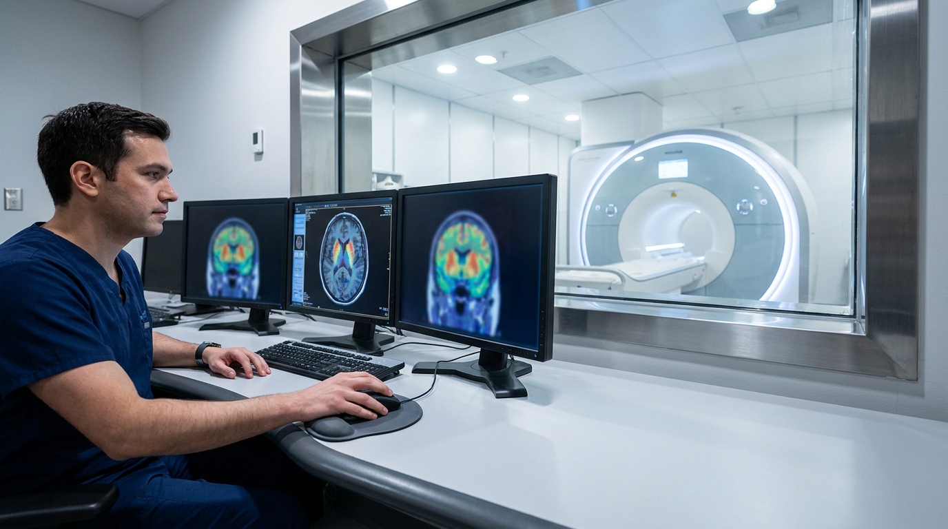
For Doctors in a Hurry
- Researchers investigated whether schizophrenia patients with brain structure resembling behavioral variant frontotemporal dementia also share neurochemical features of that neurodegenerative disease.
- This study evaluated 76 schizophrenia patients using magnetic resonance imaging and positron emission tomography to measure dopamine synthesis and iron accumulation.
- Higher dementia-like patterns correlated with lower striatal dopamine (r = -0.343, p = 0.032) and increased striatal iron (r = 0.282, p = 0.020).
- The researchers concluded these patients exhibit neurodegenerative hallmarks that contrast with the hyperdopaminergic model typically associated with schizophrenia.
- Identifying this dementia-like subtype may help clinicians recognize patients whose underlying pathology involves distinct mechanisms rather than simple dopamine dysregulation.
The Neurodegenerative Subtype of Schizophrenia
The clinical differentiation between behavioral variant frontotemporal dementia and primary psychiatric disorders remains a significant diagnostic hurdle due to profound overlaps in apathy, social withdrawal, and executive dysfunction [1, 2]. While these conditions are traditionally categorized separately, neuroimaging meta-analyses have identified shared structural alterations in the prefrontal and anterior cingulate cortices across both schizophrenia and dementia [2]. These commonalities extend to social cognitive deficits, which appear to be a core cognitive phenotype across multiple neurological and psychiatric conditions [3]. Furthermore, physiological markers such as olfactory identification impairments are increasingly recognized as potential tools to help clinicians distinguish neurodegenerative processes from primary psychiatric illness [4]. Building on this overlap, researchers recently investigated whether a specific subset of patients with schizophrenia exhibits the deep neurochemical and structural signatures typically associated with frontotemporal degeneration, raising the prospect of identifying distinct biological subtypes to guide future treatment.
Mapping the Frontotemporal Phenotype
The researchers analyzed a cohort of 76 patients with schizophrenia (mean age 31.2 years; 71% male) drawn from a previous clinical trial (NCT04038957). To establish a rigorous baseline, the study utilized a control group of 79 healthy individuals for age-related dynamic standardization, a statistical technique that accounts for normal age-linked brain changes to isolate disease-specific deviations. All 76 patients underwent structural magnetic resonance imaging (MRI) to measure gray matter volume. To identify specific neuroanatomical subtypes, the authors applied a previously validated machine-learning-based gray matter volume classifier. This algorithm quantified the degree to which each patient expressed a structural pattern characteristic of behavioral variant frontotemporal dementia (bvFTD). By using this tool, the researchers could pinpoint which patients possessed a brain structure mirroring the atrophy patterns typically seen in this specific neurodegenerative dementia. The investigation then extended beyond structural anatomy into neurochemical markers through a multimodal imaging approach. A subset of 40 patients underwent [18F]-DOPA positron emission tomography (PET) to measure striatal dopamine synthesis capacity, tracking the rate at which the brain produces this neurotransmitter. Additionally, 68 patients received neuromelanin-sensitive MRI to assess midbrain dopaminergic integrity, a measure of the health and density of dopamine-producing neurons. Finally, 69 patients were evaluated using quantitative susceptibility mapping, a specialized MRI technique used to measure brain iron accumulation. This comprehensive imaging battery allowed the team to correlate the dementia-like structural signature with specific biochemical and mineral changes in the striatum and midbrain.
Striatal Hypodopaminergia and Iron Overload
The researchers identified a distinct biochemical profile in patients who scored higher on the behavioral variant frontotemporal dementia (bvFTD) gray matter volume classifier. Specifically, higher bvFTD-like scores were significantly associated with lower striatal dopamine synthesis capacity (r = -0.343, p = 0.032). Furthermore, the study found that higher bvFTD-like scores were significantly associated with higher striatal quantitative susceptibility mapping values (r = 0.282, p = 0.020), indicating increased brain iron accumulation. Together, these findings suggest that this patient subgroup experiences both a deficit in dopamine production and an increase in iron deposition within the striatum. In contrast, these pathological changes did not appear to extend to the midbrain, pointing to a localized rather than systemic dopaminergic failure. The researchers reported no significant association between bvFTD-like scores and quantitative susceptibility mapping values in the dopaminergic midbrain (p = 0.903). Similarly, no significant association was found with neuromelanin-sensitive MRI values in the dopaminergic midbrain (p = 0.102), indicating that the structural and functional integrity of midbrain dopamine-producing neurons remains relatively preserved. Collectively, these data demonstrate that patients with schizophrenia who express a stronger bvFTD-like gray matter volume pattern exhibit lower striatal dopamine synthesis and elevated striatal iron. For the practicing clinician, this reveals that schizophrenia is not a monolithic condition driven solely by dopamine overactivity. Instead, a subset of patients possesses a neurodegenerative-like profile characterized by striatal hypodopaminergia (low dopamine levels) and iron overload. Because these are hallmark features of behavioral variant frontotemporal dementia rather than primary psychosis, patients with this phenotype may eventually require entirely different management strategies than those typically employed for classic schizophrenia.
Clinical Implications and the Hyperdopaminergic Model
The identification of this neurochemical profile suggests that the behavioral variant frontotemporal dementia-like phenotype is an intrinsic biological subtype of the disease rather than a secondary effect of clinical presentation or treatment. The researchers found no significant associations between the dementia-like gray matter pattern and negative symptom severity, indicating that the observed structural and chemical changes are not merely a proxy for apathy or social withdrawal. Furthermore, no significant associations were found with medication measures. This lack of correlation with antipsychotic exposure is highly relevant for the practicing clinician, as it suggests the observed striatal hypodopaminergia and iron accumulation are not artifacts of long-term dopamine receptor blockade, but rather represent a distinct underlying pathophysiology. These findings fundamentally contrast with the traditional hyperdopaminergic model of schizophrenia, which posits that the disorder is driven by an excess of dopamine in the striatum. By demonstrating a subgroup characterized by lower striatal dopamine synthesis and elevated striatal iron, this study challenges the universal application of that framework. Clinically, this suggests that patients expressing this specific phenotype may be less likely to respond to standard D2-receptor antagonists, which are designed to mitigate dopamine overactivity. Instead, this subgroup may eventually require therapeutic strategies that address neurodegenerative-like mechanisms or iron-mediated oxidative stress, highlighting the need for a more personalized approach to psychiatric management that accounts for this unique low-dopamine, high-iron signature.
References
1. Rascovsky K, Hodges JR, Knopman DS, et al. Sensitivity of revised diagnostic criteria for the behavioural variant of frontotemporal dementia. Brain. 2011. doi:10.1093/brain/awr179
2. Ulugut H, Trieu C, Groot C, et al. Overlap of Neuroanatomical Involvement in Frontotemporal Dementia and Primary Psychiatric Disorders: A Meta-analysis.. Biological psychiatry. 2023. doi:10.1016/j.biopsych.2022.05.028
3. Cotter J, Granger K, Backx R, Hobbs M, Looi CY, Barnett JH. Social cognitive dysfunction as a clinical marker: A systematic review of meta-analyses across 30 clinical conditions. Neuroscience & Biobehavioral Reviews. 2017. doi:10.1016/j.neubiorev.2017.11.014
4. Carnemolla SE, Hsieh JW, Sipione R, et al. Olfactory dysfunction in frontotemporal dementia and psychiatric disorders: A systematic review.. Neuroscience and biobehavioral reviews. 2020. doi:10.1016/j.neubiorev.2020.08.002