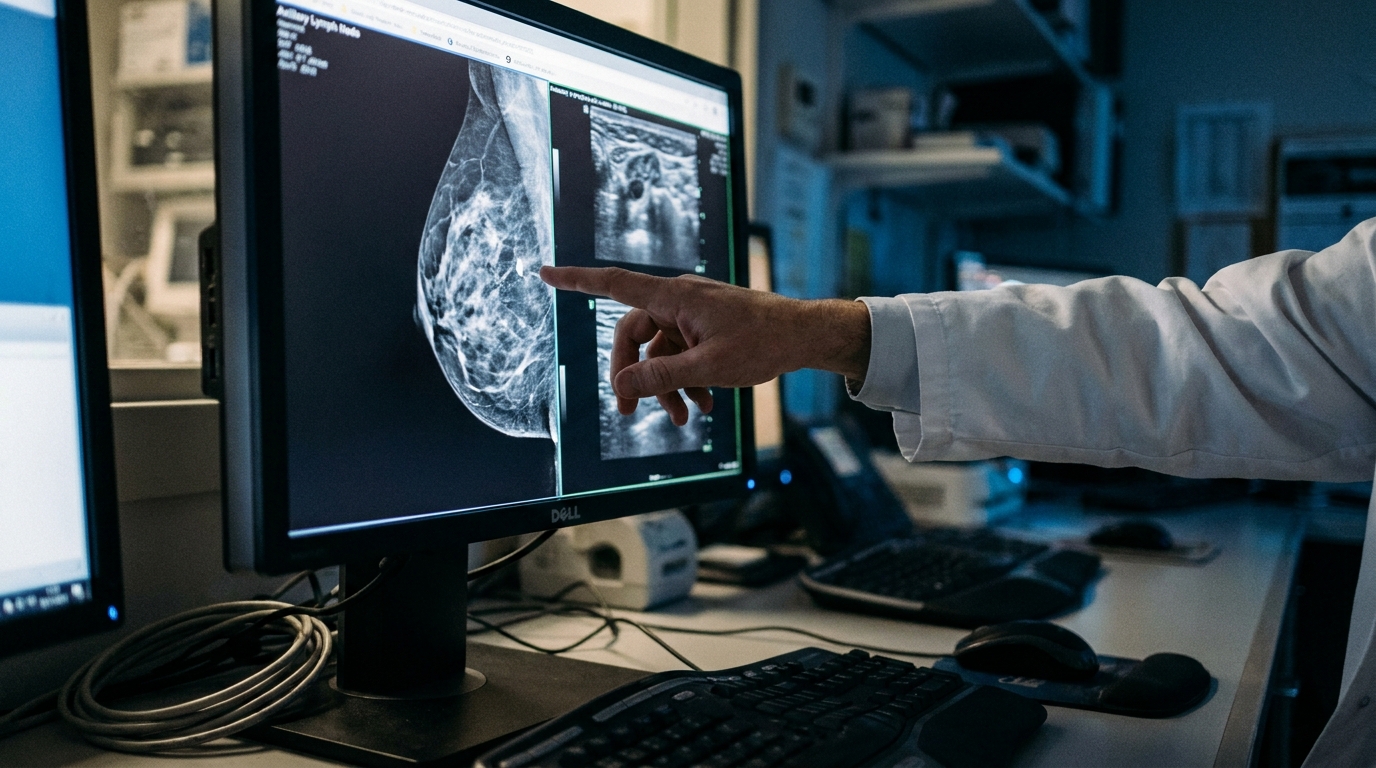
For Doctors in a Hurry
- Researchers investigated if sentinel lymph node biopsy could safely replace axillary dissection in patients with clinically node-positive, hormone receptor-positive breast cancer.
- This nonrandomized trial enrolled 78 patients with early-stage disease and three or fewer abnormal nodes identified by axillary ultrasound.
- Sentinel lymph node biopsy alone was performed in 76 percent of patients, while 24 percent required axillary lymph node dissection.
- The researchers concluded that this approach is feasible for node-positive disease when combined with the resection of palpable nodes.
- This strategy potentially reduces surgical morbidity by omitting extensive axillary dissection in patients with a limited nodal burden.
De-escalating Axillary Surgery in Node-Positive Breast Cancer
The management of the axilla in early-stage breast cancer has shifted significantly toward de-escalation to minimize the high morbidity associated with complete axillary lymph node dissection [1, 2]. While omitting axillary dissection is now standard for clinically node-negative patients with limited sentinel lymph node involvement, those presenting with palpable adenopathy have historically been excluded from these conservative protocols [3, 4]. Complete dissection remains the default for these patients, despite its well-documented risks of lymphedema, sensory loss, and reduced range of motion [5, 6]. Recent meta-analyses suggest that survival outcomes may not be compromised by less invasive approaches in select populations, yet clinicians lack prospective data for those with palpable disease [7, 8]. A new study now evaluates whether upfront sentinel lymph node biopsy can be successfully applied to this higher-risk cohort to further reduce surgical overtreatment.
Trial Design and Patient Selection Criteria
To evaluate the feasibility of upfront sentinel lymph node biopsy in patients traditionally managed with immediate complete axillary dissection, researchers conducted a multicenter, nonrandomized clinical trial. The study enrolled participants with hormone receptor-positive, human epidermal growth factor receptor 2-negative (HR+/HER2-) breast cancer, the most common breast cancer subtype, staged as cTx/cT1-2 cN1. To be eligible, patients had to present with palpable, mobile level I or II axillary adenopathy but demonstrate limited nodal involvement on axillary ultrasound, defined as having three or fewer morphologically abnormal nodes. This prospective trial was conducted across four specialized centers and began on April 20, 2021, with the final database frozen for analysis on September 26, 2024. Preoperative axillary ultrasound served as the primary tool for patient selection and risk stratification, allowing clinicians to noninvasively estimate nodal burden before surgery. Among the enrolled cohort, 39 patients (50%) presented with a single abnormal-appearing node on ultrasound, while 33 patients (42%) had two abnormal nodes, and 6 patients (8%) had three. Following this imaging assessment, all participants underwent upfront surgical intervention consisting of either a lumpectomy or mastectomy. This was combined with a sentinel lymph node biopsy using single or dual-tracer mapping (the use of radioactive technetium, blue dye, or both to identify the primary draining nodes). This surgical strategy aimed to identify patients with a low nodal burden who might safely avoid the morbidity of a full axillary lymph node dissection.
Clinical and Pathological Characteristics
The study cohort consisted of 78 enrolled patients with a median age of 58 years (interquartile range, 49.0 to 66.5 years). Clinical staging at the time of enrollment showed that nearly all participants had early-stage invasive disease. Specifically, 37 patients (47%) presented with cT1 tumors, while 40 patients (51%) were staged as cT2. Pathological analysis of the primary tumors revealed that 56 patients (72%) had ductal histology, and the majority of the cohort, 59 tumors (76%), were moderately differentiated. The median pathologic tumor size was 2.3 cm (interquartile range, 1.6 to 3.3 cm). These findings provide a detailed profile of the HR+/HER2- population studied, highlighting that the tumors were of sufficient size and grade to warrant careful axillary evaluation. Despite the relatively small tumor sizes, the cohort exhibited several high-risk pathological features associated with nodal involvement. Lymphovascular invasion (the presence of cancer cells within the lumen of lymphatic or blood vessels) was identified in 50 patients (64%). Furthermore, extracapsular extension (the spread of tumor cells beyond the lymph node capsule into the surrounding adipose tissue, a feature historically prompting aggressive surgery) was present in 54 patients (69%). The high prevalence of these features underscores the clinical challenge of managing cN1 disease, as these markers often correlate with a higher burden of axillary disease.
Surgical Feasibility and Nodal Mapping Success
The technical execution of the sentinel lymph node biopsy proved highly successful within this cohort of patients with palpable axillary disease. To maximize the detection of the sentinel nodes, the researchers utilized a rigorous mapping protocol. Specifically, sentinel lymph node biopsy was performed with dual tracer in 68 patients (87%), employing both radioactive colloid and blue dye to identify the lymphatic drainage from the primary tumor. This approach facilitated a high degree of surgical precision, as the researchers reported that 3 or more sentinel lymph nodes were retrieved in 75 patients (96%). This high retrieval rate is a critical metric for surgeons, as the removal of at least three nodes is traditionally associated with a lower false-negative rate and a more accurate assessment of the total nodal burden in the axilla. However, a key finding regarding the identification of diseased nodes suggests that relying solely on tracers may be insufficient in the cN1 setting. The study found that palpable diseased nodes were blue, radioactive, or both in only 107 of 161 instances (66.5%). This means that roughly one-third of the clinically suspicious nodes did not take up the mapping agents. This discrepancy underscores the necessity of the surgeon manually identifying and resecting all palpable nodes during the procedure, regardless of whether they exhibit tracer activity. By ensuring the removal of both the sentinel nodes and any clinically abnormal nodes, the surgical team can more reliably identify patients with limited nodal involvement who may safely bypass a full axillary lymph node dissection.
Nodal Burden and Axillary Dissection Rates
The primary outcome of this clinical trial was the rate of axillary lymph node dissection, a procedure traditionally mandated for patients with palpable nodal disease. Upon surgical evaluation of the 78 enrolled patients, the researchers characterized the pathologic nodal burden to determine who required more extensive surgery. The analysis revealed that 24 patients (31%) had 1 positive sentinel lymph node, while 30 patients (38%) had 2 positive sentinel lymph nodes. Combined, these groups represent a significant majority of the cohort who presented with limited nodal involvement despite having clinically palpable disease at baseline. In contrast, 24 patients (31%) were found to have 3 or more positive sentinel lymph nodes, meeting the study threshold for more extensive surgical intervention. Based on the trial protocol, axillary lymph node dissection was indicated if three or more positive sentinel lymph nodes were identified during the upfront procedure. Following these criteria, sentinel lymph node biopsy alone was performed in 59 patients (76%), allowing the majority of the study population to avoid the morbidity associated with full nodal clearance. Conversely, 19 patients (24%) underwent axillary lymph node dissection due to their higher nodal burden. The researchers also noted that indicated axillary lymph node dissection was deferred in 5 cases. For practicing oncologists, these dissection rates illustrate a tangible clinical shift toward surgical de-escalation in patients with HR+/HER2- disease who demonstrate a limited number of positive nodes upon pathologic review.
Early Locoregional Control and Clinical Implications
The researchers established secondary outcomes to evaluate the oncologic safety and technical precision of this de-escalated surgical approach, specifically tracking the rate of locoregional recurrence. Clinical follow-up data suggest that omitting axillary lymph node dissection does not compromise early disease control in this specific patient population. Among 68 patients with 12 months or more of follow-up (median follow-up of 25 months), there were no isolated axillary or locoregional recurrences. For the practicing clinician, these results indicate that upfront sentinel lymph node biopsy is a feasible and safe strategy for patients with HR+/HER2- cN1 breast cancer who have three or fewer morphologically abnormal nodes on ultrasound. The data also reinforce a critical surgical caveat: physical resection of palpable nodes is necessary to minimize false-negative rates, as relying solely on tracers missed 33.5% of diseased nodes in this cohort. Ultimately, by identifying the 76% of patients who have a limited nodal burden, surgeons can safely avoid full axillary dissection and its associated long-term morbidity (such as lymphedema and neuropathy) without sacrificing early regional stability.
References
1. Bakri NAC, Kwasnicki RM, Khan N, et al. Impact of Axillary Lymph Node Dissection and Sentinel Lymph Node Biopsy on Upper Limb Morbidity in Breast Cancer Patients: A Systematic Review and Meta-Analysis.. Annals of surgery. 2023. doi:10.1097/SLA.0000000000005671
2. Yan M, Abdi MA, Falkson C. Axillary Management in Breast Cancer Patients: A Comprehensive Review of the Key Trials.. Clinical breast cancer. 2018. doi:10.1016/j.clbc.2018.08.002
3. Lyman GH, Giuliano AE, Somerfield MR, et al. American Society of Clinical Oncology Guideline Recommendations for Sentinel Lymph Node Biopsy in Early-Stage Breast Cancer. Journal of Clinical Oncology. 2005. doi:10.1200/jco.2005.08.001
4. Lyman GH, Temin S, Edge SB, et al. Sentinel Lymph Node Biopsy for Patients With Early-Stage Breast Cancer: American Society of Clinical Oncology Clinical Practice Guideline Update. Journal of Clinical Oncology. 2014. doi:10.1200/jco.2013.54.1177
5. Pilger TL, Francisco DF, Reis FJCD. Effect of sentinel lymph node biopsy on upper limb function in women with early breast cancer: A systematic review of clinical trials.. European journal of surgical oncology : the journal of the European Society of Surgical Oncology and the British Association of Surgical Oncology. 2021. doi:10.1016/j.ejso.2021.01.024
6. Mansel RE, Fallowfield L, Kissin M, et al. Randomized Multicenter Trial of Sentinel Node Biopsy Versus Standard Axillary Treatment in Operable Breast Cancer: The ALMANAC Trial. JNCI Journal of the National Cancer Institute. 2006. doi:10.1093/jnci/djj158
7. Agrawal SK, Imran MM, Sethi S, Yadav SK, Agarwal P, Sharma D. Sentinel Lymph Node Biopsy vs. Axillary Lymph Node Dissection for Early-Stage Breast Cancer and Sentinel Lymph Node Metastasis: An Updated Systematic Review and Meta-Analysis With Special Focus on Locoregional Recurrence and Regional Node Irradiation.. Breast cancer : basic and clinical research. 2025. doi:10.1177/11782234251335409
8. Li C, Zhang P, Lv J, et al. Axillary management in patients with clinical node-negative early breast cancer and positive sentinel lymph node: a systematic review and meta-analysis.. Frontiers in oncology. 2023. doi:10.3389/fonc.2023.1320867