
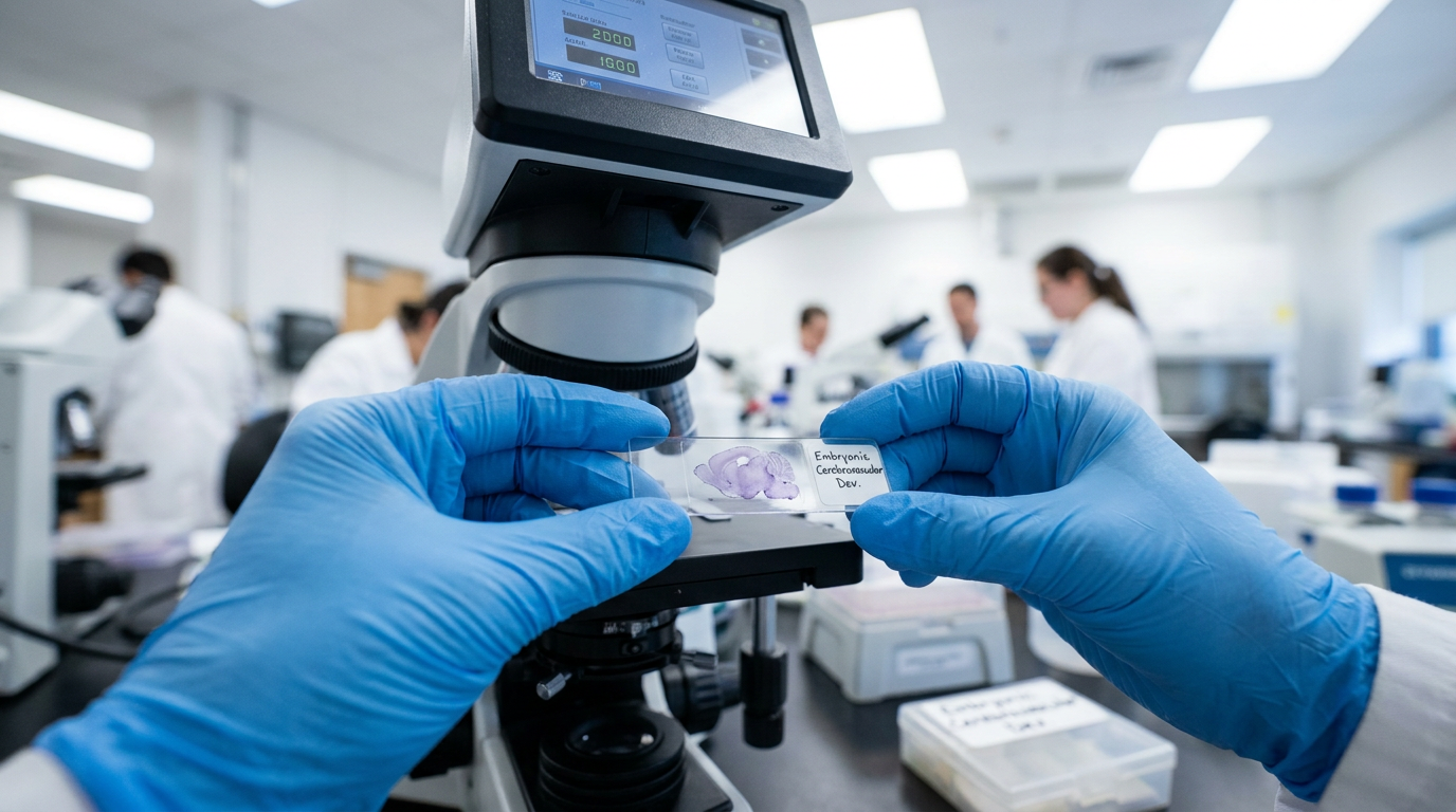
- The study investigated how tissue factor pathway inhibitor (TFPI) deficiency causes embryonic lethality and cerebrovascular defects.
- Researchers bred a hyperactivatable mouse protein C (hMPC) transgene into TFPI-deficient mice, observing adult survival.
- The hMPC transgene reduced glomeruloid body numbers by only 36% but resolved them by postnatal day 10.
- The authors concluded TFPI is an essential, temporally acting inhibitor of thrombin generation during embryonic cerebral angiogenesis.
- These findings underscore the critical role of coagulation proteases in diverse biological processes beyond hemostasis.
Coagulation's Unseen Hand in Brain Vascular Development
The integrity of the cerebrovascular system is essential for neurological function, and its disruption during development can have lasting consequences [1, 2]. Angiogenesis, the formation of new blood vessels, is a tightly regulated process that, when dysregulated, contributes to pathologies ranging from aggressive cancers like glioblastoma to thrombotic microangiopathies [3, 4, 5, 6, 7]. While the function of coagulation factors in hemostasis is well-established, their specific contributions to embryonic vascular development remain an area of active investigation [8]. A recent preclinical study provides new insight into this relationship, demonstrating how a key anticoagulant protein is indispensable for the proper formation of the brain's vascular architecture, a finding with potential relevance for understanding certain congenital vascular defects [9].
TFPI Deficiency: A Fatal Flaw in Cerebrovascular Formation
A preclinical mouse model has illuminated the indispensable role of tissue factor pathway inhibitor (TFPI), a primary physiological regulator of the extrinsic coagulation pathway. The study demonstrated that a complete absence of this protein is incompatible with life, as mice genetically engineered to lack TFPI (Tfpi-/-) succumbed to embryonic lethality. This fatal outcome was attributed to excess thrombin production and the formation of severe cerebrovascular defects, which the researchers termed glomeruloid bodies. These glomeruloid bodies are tangled, malformed clusters of blood vessels, pathologically similar to structures observed in some human conditions like glioblastoma. The findings establish that without TFPI's inhibitory function, the uncontrolled activity of thrombin during a critical developmental window disrupts the normal process of brain angiogenesis, leading to these catastrophic structural failures and preventing survival to term.
Hyperactivatable Protein C: A Partial Rescue
To determine if another potent anticoagulant could compensate for the absence of TFPI, the researchers introduced a transgene that produces a hyperactivatable form of mouse protein C (hMPC). This engineered protein provides enhanced anticoagulant and cytoprotective activity. When this transgene was expressed in TFPI-deficient embryos, the study found that Tfpi-/-/hMPC+ embryos survived to adulthood, a complete reversal of the embryonic lethality. This outcome demonstrates that augmenting the protein C pathway can overcome the immediate fatal consequences of unchecked thrombin generation during development. However, this functional rescue was not accompanied by a complete structural correction. The hMPC transgene reduced the number of glomeruloid bodies by only 36%. Furthermore, it did not prevent fibrin deposition or the disruption of the blood-brain barrier within the remaining vascular malformations, indicating that while survival was achieved, the underlying anatomical defects persisted into the embryonic period.
Beyond Structural Repair: Cytoprotective Effects and Temporal Resolution
The survival of TFPI-deficient embryos, despite persistent vascular malformations, pointed toward mechanisms beyond structural repair. The researchers observed decreased hypoxia and reduced cellular death in the brains of the rescued Tfpi-/-/hMPC+ mice. This suggests that the cytoprotective effects of hyperactivatable protein C, which are known to be distinct from its anticoagulant functions, contributed significantly to the rescue. By mitigating cellular stress and injury, hMPC allowed the embryos to survive even with a partially compromised cerebral vasculature. In a remarkable long-term observation, the study also revealed a distinct temporal dynamic. Although present at birth, the glomeruloid bodies were completely resolved in Tfpi-/-/hMPC+ pups by postnatal day 10. This finding indicates that while TFPI is essential to prevent the initial formation of these defects during embryonic life, the brain possesses a robust capacity for postnatal vascular remodeling and repair when supported by the enhanced activity of the protein C pathway.
Angiogenesis Dysregulation: The Molecular Signature of TFPI Deficiency
To define the molecular drivers of these vascular defects, the investigators performed bulk RNA sequencing, a technique that provides a comprehensive snapshot of gene activity across a tissue sample. Analysis of brain tissue from embryonic day 15.5 identified increased angiogenesis as the overwhelming biological process altered in the Tfpi-/- brain. This genetic evidence confirms that the absence of TFPI leads to uncontrolled blood vessel growth. Specifically, the analysis revealed altered expression of genes critical to vessel formation, including apelin (a peptide that promotes vessel sprouting), adrenomedulin (a factor involved in vasodilation and angiogenesis), and UNC5b (a receptor that guides developing vessels). This molecular signature was consistent with the histological finding of abundant endothelial tip cells, the specialized cells that lead the formation of new blood vessels, within the glomeruloid bodies. Critically, the study showed that the increased expression of these pro-angiogenic genes was reversed by the hMPC transgene, directly linking the therapeutic intervention to the normalization of the underlying genetic program driving the pathology.
Clinical Implications: TFPI's Unique Role in Brain Development
This preclinical research establishes that TFPI is an essential and non-redundant inhibitor of thrombin generation during embryonic brain angiogenesis. Its function is highly specific, acting temporally within or around the developing cerebral vasculature to ensure proper vessel formation. A key clinical insight is that this role cannot be fully compensated for by other powerful anticoagulants. While enhancing protein C activity rescued lethality through cytoprotective mechanisms and enabled eventual postnatal repair, it did not prevent the initial embryonic vascular injury. This dissociation underscores a critical concept for clinicians: coagulation proteases and their inhibitors have highly specialized roles in organogenesis that extend far beyond their classical functions in hemostasis. This work deepens our understanding of the fundamental biology of cerebrovascular development and may inform future investigations into congenital vascular anomalies and other conditions characterized by aberrant angiogenesis.
References
1. Ding J, Lee S, Vlahos L, et al. Therapeutic blood-brain barrier modulation and stroke treatment by a bioengineered FZD4-selective WNT surrogate in mice. Nature Communications. 2023. doi:10.1038/s41467-023-37689-1
2. Liebner S, Dijkhuizen RM, Reiss Y, Plate KH, Agalliu D, Constantin G. Functional morphology of the blood–brain barrier in health and disease. Acta Neuropathologica. 2018. doi:10.1007/s00401-018-1815-1
3. Allford S, Hunt BJ, Rose P, Machin SJ. Guidelines on the diagnosis and management of the thrombotic microangiopathic haemolytic anaemias. British Journal of Haematology. 2003. doi:10.1046/j.1365-2141.2003.04049.x
4. Opławski M, Michalski M, Witek A, et al. Identification of a gene expression profile associated with the regulation of angiogenesis in endometrial cancer. Molecular Medicine Reports. 2017. doi:10.3892/mmr.2017.6868
5. Stefansson IM, Ræder MB, Wik E, et al. Increased angiogenesis is associated with a 32-gene expression signature and 6p21 amplification in aggressive endometrial cancer. Oncotarget. 2015. doi:10.18632/oncotarget.3521
6. Ballen AIC, Amosu M, Ravinder S, et al. Metabolic Reprogramming in Glioblastoma Multiforme: A Review of Pathways and Therapeutic Targets. Cells. 2024. doi:10.3390/cells13181574
7. Bae E, Huang P, Müller-Greven G, et al. Integrin α3β1 promotes vessel formation of glioblastoma-associated endothelial cells through calcium-mediated macropinocytosis and lysosomal exocytosis. Nature Communications. 2022. doi:10.1038/s41467-022-31981-2
8. Pablo-Moreno JAD, Serrano LJ, Revuelta L, Sánchez MJ, Liras A. The Vascular Endothelium and Coagulation: Homeostasis, Disease, and Treatment, with a Focus on the Von Willebrand Factor and Factors VIII and V. International Journal of Molecular Sciences. 2022. doi:10.3390/ijms23158283
9. Maroney SA, Martinez ND, Krishnamoorthy P, et al. Defective cerebrovascular development in mice lacking TFPI is restored by activated protein C. Blood. 2026. doi:10.1182/blood.2025031420