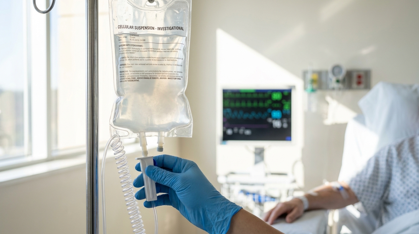
For Doctors in a Hurry
- Clinicians lack effective treatments for the chronic neuroinflammation and progressive tissue loss that follow traumatic brain injury.
- This Phase 1/2a study evaluated three intravenous infusions of autologous adipose-derived mesenchymal stromal cells in 24 patients.
- Treatment significantly reduced hippocampal mean diffusivity (a measure of tissue water diffusion) by 163.51 cubic millimeters (p=0.013).
- The authors observed significant reductions in brain immune cell density and improvements in patient-reported depression, fatigue, and pain (p<0.03).
- These data provide a quantitative basis for larger clinical trials targeting functional recovery and surrogate imaging markers in chronic injury.
Addressing the Chronic Inflammatory Cascade in Traumatic Brain Injury
Traumatic brain injury often evolves from an acute mechanical event into a progressive, chronic condition marked by persistent neuroinflammation and subsequent neurodegeneration [1, 2]. While acute care focuses on immediate stabilization, survivors frequently face lasting neurological deficits with no therapies available to reverse established damage [3]. One potential strategy involves mesenchymal stromal cells, which are multipotent progenitor cells capable of modulating the immune environment and releasing neuroprotective factors [4, 5]. Although their use in related fields like spinal cord injury has raised safety questions [6, 7], a recent Phase 1/2a study in 24 patients with chronic TBI offers new data. The findings show that three intravenous infusions of autologous adipose-derived mesenchymal stromal cells significantly reduced brain immune cell density in the right caudate (p < 0.0003) and improved microstructural integrity in the hippocampus (p = 0.013), as measured by decreased mean diffusivity volumes [8].
Safety and Protocol of Autologous Stromal Cell Infusion
The Phase 1/2a trial evaluated a cellular therapy in 24 patients with chronic traumatic brain injury. The protocol involved harvesting mesenchymal stromal cells from each patient's own adipose tissue, an autologous approach designed to eliminate the risk of immunological rejection. Each participant then received three intravenous infusions of these autologous cells, known as HB-adMSCs, over a six-week period. This schedule was intended to provide sustained exposure to the cells' immunomodulatory effects during the chronic phase of injury. Safety was a primary endpoint, and the study confirmed the protocol was well-tolerated, reporting that there were no serious treatment-related adverse events among the participants. This finding is clinically important, as it helps address concerns about potential embolic or systemic inflammatory risks associated with intravascular cell delivery. Beyond safety, the study demonstrated that the therapy produced measurable effects on both functional and neurocognitive outcomes, providing the quantitative data needed to properly power future, larger clinical trials using either clinical recovery or surrogate imaging markers as endpoints.
Quantifying Reductions in Brain Immune Cell Density
To directly measure the therapy's effect on neuroinflammation, the researchers employed positron emission tomography (PET) with the radiotracer [11C]ER176. This imaging technique allows for in-vivo quantification of brain immune cell density, providing an objective marker of the microglial activation that drives the chronic inflammatory state in TBI. The results demonstrated that the treatment induced significant reductions in brain immune cell density in several key brain regions. The most substantial decrease was observed in the right caudate (x,y,z: 12,2,7; T20 = 4.1; P < 0.0003), a structure involved in motor control and learning. This effect extended into the adjacent right ventral anterior thalamus (x,y,z: 13, -2,11; T20 = 3.6; P < 0.0009), which relays sensory and motor signals, and the right nucleus accumbens (x,y,z: 8,9, -3; T20 = 2.5; P < 0.01), a central component of the brain's reward circuitry. A significant reduction was also noted in the right parahippocampal gyrus (x,y,z: 29, -40, -7; T20 = 3.1; P < 0.003), a region critical for memory encoding. These anatomical findings provide a biological correlate for the clinical improvements seen in patients, as these brain regions are functionally linked to the regulation of pain, fatigue, and mood.
Improvements in Microstructural White Matter Integrity
The study also assessed changes in brain structure using diffusion tensor MRI (DT-MRI), an imaging modality that evaluates the integrity of white matter by measuring the diffusion of water molecules. In damaged or disorganized tissue, water diffuses more freely, resulting in elevated mean diffusivity. The researchers quantified the volume of tissue with abnormally high water diffusion, a metric they termed supraMD, to track microstructural changes. Six months after treatment, they found that elevated mean diffusivity volumes were significantly reduced bilaterally in the hippocampus, a region vital for memory, with a mean decrease of 163.51 mm3 (95% CI -286.55 to -41.51 mm3; P=0.013). This suggests a repair or stabilization of white matter tracts. A similar trend was observed in the amygdala, which is involved in emotional processing, with a mean supraMD decrease of 113.60 mm3 (95% CI -229.52 to 2.90 mm3; P=0.058). No significant changes were seen in the insula (P=0.19). Importantly, while these microstructural measures improved, macrostructural volumetric analyses showed no significant changes in the size of the hippocampus, amygdala, or insula (all P > 0.10). This distinction suggests the therapy may improve the quality and organization of existing neural tissue rather than generating new brain volume.
Clinical Correlation with Mood and Somatic Symptoms
The biological changes observed on imaging corresponded with tangible clinical benefits for patients. Six months after the final infusion, participants reported significant reductions in measures of depression (P=0.026), fatigue (P=0.008), and pain (P=0.007). These symptomatic improvements align with the observed decreases in immune cell density within brain regions that regulate mood, arousal, and sensory processing. Furthermore, the study uncovered a direct link between patients' baseline psychological state and their neurobiological response to the therapy. The researchers found significant interactions between baseline levels of anxiety and depression and the magnitude of microstructural improvement (reduction in supraMD) in the amygdala and hippocampus, respectively. This suggests that a patient's initial clinical profile may predict their capacity for structural repair following this cellular intervention. By linking functional recovery to objective measures of reduced neuroinflammation and improved white matter integrity, the study provides a strong rationale for advancing autologous mesenchymal stromal cell therapy into later-phase clinical trials for chronic TBI.
References
1. Alizada M, Lin S, Gao H, et al. Cellular therapy for traumatic brain injury in adults: a meta-analysis of controlled clinical trials. Archives of Medical Science. 2024. doi:10.5114/aoms/194535
2. Pischiutta F, Caruso E, Lugo A, et al. Systematic review and meta-analysis of preclinical studies testing mesenchymal stromal cells for traumatic brain injury.. NPJ Regenerative medicine. 2021. doi:10.1038/s41536-021-00182-8
3. Bonilla C, Zurita M. Cell-Based Therapies for Traumatic Brain Injury: Therapeutic Treatments and Clinical Trials. Biomedicines. 2021. doi:10.3390/biomedicines9060669
4. Zanier ER, Pischiutta F, Rulli E, et al. MesenchymAl stromal cells for Traumatic bRain Injury (MATRIx): a study protocol for a multicenter, double-blind, randomised, placebo-controlled phase II trial.. Intensive care medicine experimental. 2023. doi:10.1186/s40635-023-00535-1
5. Hoang DM, Pham PT, Bach TQ, et al. Stem cell-based therapy for human diseases. Signal Transduction and Targeted Therapy. 2022. doi:10.1038/s41392-022-01134-4
6. Lalu MM, McIntyre L, Pugliese C, et al. Safety of Cell Therapy with Mesenchymal Stromal Cells (SafeCell): A Systematic Review and Meta-Analysis of Clinical Trials. PLoS ONE. 2012. doi:10.1371/journal.pone.0047559
7. Shang Z, Wang M, Zhang B, Wang X, Wanyan P. Clinical translation of stem cell therapy for spinal cord injury still premature: results from a single-arm meta-analysis based on 62 clinical trials. BMC Medicine. 2022. doi:10.1186/s12916-022-02482-2
8. Cox CS, Ashley JR, Juranek J, et al. Autologous adipose-derived mesenchymal stromal cells for chronic traumatic brain injury.. Brain : a journal of neurology. 2026. doi:10.1093/brain/awag165