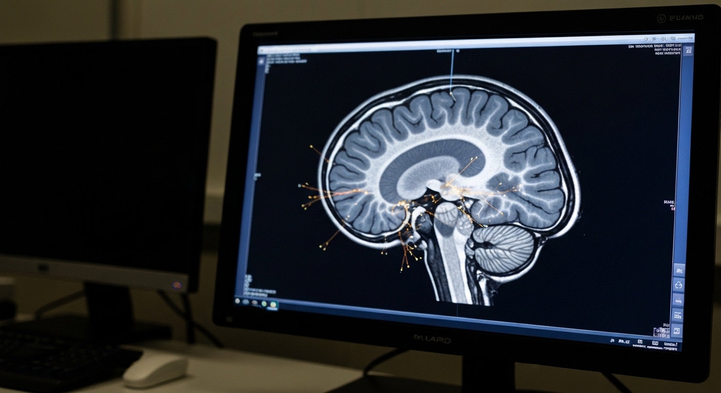
For Doctors in a Hurry
- Clinicians lack clarity on whether transcranial magnetic stimulation induces target-specific connectivity changes that correlate with improved patient outcomes.
- The researchers randomized thirty-six patients with depression and anxiety to receive thirty sessions of transcranial magnetic stimulation at two distinct targets.
- Stronger baseline connectivity to the targeted circuit predicted greater connectivity changes and anxiety improvement with a correlation of 0.47, p=0.004.
- The authors conclude that baseline connectivity serves as a more reliable biomarker for treatment response than post-treatment connectivity changes.
- Physicians should consider individual circuit architecture when defining targeting protocols to improve clinical outcomes for patients with anxiety symptoms.
Refining Neural Targets in Treatment Resistant Depression
Transcranial magnetic stimulation (TMS) has become a standard intervention for patients with major depressive disorder who have failed multiple antidepressant trials [1]. Beyond its established role in treating core depressive symptoms, meta-analytic evidence supports its efficacy in addressing comorbid pathological anxiety and obsessive-compulsive symptoms [2, 3]. Clinical protocols have traditionally focused on the dorsolateral prefrontal cortex, yet researchers are increasingly exploring the dorsomedial prefrontal cortex as a potential target for specific symptom dimensions like anxiety [4, 5]. Despite these advances, the heterogeneity of patient response remains a significant challenge, leading to a search for biomarkers that can predict which individuals will achieve remission [6]. A new study investigates whether these clinical improvements are truly driven by specific changes in the targeted neural circuits or if other physiological factors are at play.
Targeted circuit engagement remains the primary goal of modern neuromodulation, yet the relationship between specific stimulation sites and clinical outcomes requires further validation. This head-to-head clinical trial enrolled 36 patients diagnosed with comorbid depression and anxiety, randomizing them to receive 30 sessions of transcranial magnetic stimulation (TMS). The researchers sought to determine if stimulating specific prefrontal regions would yield distinct clinical improvements based on the patient's symptom profile. Clinical outcomes and symptom severity were rigorously assessed using two standardized scales: the Beck Depression Inventory and the Beck Anxiety Inventory. The researchers investigated two distinct neuroanatomical sites to test the specificity of treatment response. One group received stimulation to the dorsolateral prefrontal cortex, a target hypothesized to specifically improve dysphoric symptoms, such as low mood and hopelessness. The second group was randomized to the dorsomedial prefrontal cortex, which the authors hypothesized would better address anxiosomatic symptoms, including physical manifestations of anxiety like tension or autonomic arousal. While 36 patients began the trial, the final analysis focused on 29 participants who provided complete datasets, including both pre-treatment and post-treatment MRI scans. This allowed the team to correlate clinical changes with objective measures of neural connectivity, specifically looking at how the stimulation engaged the intended brain circuits.
Dissociation Between Connectivity Changes and Clinical Response
Physiological modulation of brain networks does not always translate to symptom relief, a finding that challenges current assumptions about the mechanism of TMS. To evaluate the physiological impact of the intervention, the researchers defined target engagement as the change in average functional connectivity (a measure representing the synchronized activity between distinct brain regions) within each pre-specified circuit. Following the 30-session treatment course, neuroimaging data revealed that functional connectivity decreased within both the dysphoric and anxiosomatic circuits. However, these neural shifts lacked the anatomical specificity the researchers had hypothesized. The connectivity changes following stimulation were not clearly specific to the assigned target group, meaning that stimulating the dorsolateral prefrontal cortex did not exclusively or preferentially alter its corresponding dysphoric circuit compared to the effects observed in the dorsomedial group. The study further examined whether these physiological alterations translated into measurable patient benefits using Spearman's partial correlation (a statistical method used to measure the strength of a relationship between two variables while accounting for the influence of other factors) to assess associations while controlling for relevant covariates. A critical finding emerged from this analysis: TMS-induced connectivity changes did not predict clinical symptom improvement on either the Beck Depression Inventory or the Beck Anxiety Inventory. While the stimulation successfully modulated the synchronized firing patterns within the targeted prefrontal networks, the magnitude of these post-treatment connectivity changes failed to correlate with the degree of relief patients experienced from their depressive or anxious symptoms. This suggests that the mere induction of connectivity changes may not be the primary driver of clinical recovery in this therapeutic context.
Baseline Architecture as a Predictor of Anxiety Reduction
Pre-treatment neural architecture may be a more reliable predictor of clinical success than the treatment-induced changes themselves. While post-treatment changes in neural firing patterns did not correlate with clinical outcomes, the researchers identified that the pre-treatment state of the brain served as a robust indicator of therapeutic response. Specifically, stronger baseline connectivity at the transcranial magnetic stimulation site to the targeted circuit predicted greater connectivity change (r = -0.61, p < 0.001). This finding suggests that the initial functional integrity of the neural pathway, or the pre-existing strength of communication between the stimulation site and the target network, determines the degree to which the intervention can modulate that circuit. Beyond physiological shifts, this baseline architecture also served as a clinical marker: stronger baseline connectivity at the stimulation site to the targeted circuit predicted greater anxiety improvement (rw = 0.47, p = 0.004). For the practicing clinician, these data indicate that the success of the intervention may depend more on the patient's inherent neural architecture than on the magnitude of connectivity shifts induced during the treatment course. The analysis further revealed that these predictive effects were not uniform across all participants but were driven primarily by the anxiosomatic target group, which received stimulation at the dorsomedial prefrontal site. In contrast, the dysphoric target group did not exhibit the same clear relationship between baseline connectivity and clinical recovery. To understand this discrepancy, the researchers conducted post-hoc analyses, which suggested greater inter-individual variability in the topography of the dysphoric circuit. In this context, topography refers to the specific spatial arrangement and anatomical mapping of neural networks within an individual's brain. Because the spatial layout of the dysphoric circuit varied significantly from one patient to another, a standardized stimulation target may have been less effective at consistently engaging the intended network. This high degree of anatomical variation suggests that patients with predominant dysphoric symptoms may require more personalized targeting strategies to account for individual differences in brain architecture.
Clinical Implications for Personalized TMS Protocols
The transition from standardized to personalized neuromodulation may be necessary to overcome the limitations of current TMS protocols. The results of this trial involving 29 participants with complete neuroimaging data suggest that baseline transcranial magnetic stimulation site connectivity is a more informative biomarker than post-treatment connectivity changes. While clinicians often monitor physiological shifts following a 30-session treatment course to gauge neural engagement, these findings indicate that the initial state of the neural network is the superior predictor of clinical success. Although connectivity decreased within both the dorsolateral and dorsomedial prefrontal circuits following treatment, these changes were not specific to the stimulation target and did not correlate with symptom improvement. Instead, the pre-existing functional integrity of the circuit determined the degree of therapeutic response, with stronger baseline connectivity predicting both greater connectivity change (r = -0.61, p < 0.001) and significant anxiety reduction (rw = 0.47, p = 0.004). For the practicing physician, these data suggest that circuit architecture should be considered when defining targeting protocols to optimize outcomes in patients with depression and anxiety. The predictive value of baseline connectivity was driven primarily by the anxiosomatic target group, whereas the dysphoric target group showed more variable results. Post-hoc analyses indicated that this discrepancy may be due to greater inter-individual variability in the topography, or the specific spatial arrangement and anatomical mapping, of the dysphoric circuit. Because the spatial layout of these neural networks varies significantly between individuals, a standardized stimulation site may not consistently engage the intended circuit. These findings suggest that clinical efficacy may depend on transitioning from standardized placement to personalized protocols that account for the unique neural architecture of each patient's brain before treatment begins.
References
1. Vida RG, Sághy E, Bella R, et al. Efficacy of repetitive transcranial magnetic stimulation (rTMS) adjunctive therapy for major depressive disorder (MDD) after two antidepressant treatment failures: meta-analysis of randomized sham-controlled trials.. BMC psychiatry. 2023. doi:10.1186/s12888-023-05033-y
2. Hyde J, Carr H, Kelley NJ, et al. Efficacy of neurostimulation across mental disorders: systematic review and meta-analysis of 208 randomized controlled trials. Molecular Psychiatry. 2022. doi:10.1038/s41380-022-01524-8
3. Gay F, Singier A, Aouizerate B, Salvo F, Bienvenu T. Neuromodulation Treatments of Pathological Anxiety in Anxiety Disorders, Stressor-Related Disorders, and Major Depressive Disorder: A Dimensional Systematic Review and Meta-Analysis. Frontiers in Psychiatry. 2022. doi:10.3389/fpsyt.2022.910897
4. Jin J, Wang Y, Liang S, et al. Effects of individualized rTMS on functional connectivity related to the default mode network and frontal-parietal network in major depressive disorder: exploratory analysis of a randomized controlled trial.. NeuroImage. Clinical. 2025. doi:10.1016/j.nicl.2025.103902
5. Marques RC, Vieira L, Marques D, Cantilino A. Transcranial magnetic stimulation of the medial prefrontal cortex for psychiatric disorders: a systematic review. Revista Brasileira de Psiquiatria. 2019. doi:10.1590/1516-4446-2019-0344
6. Cao X, Deng C, Su X, Guo Y. Response and Remission Rates Following High-Frequency vs. Low-Frequency Repetitive Transcranial Magnetic Stimulation (rTMS) Over Right DLPFC for Treating Major Depressive Disorder (MDD): A Meta-Analysis of Randomized, Double-Blind Trials.. Frontiers in psychiatry. 2018. doi:10.3389/fpsyt.2018.00413