
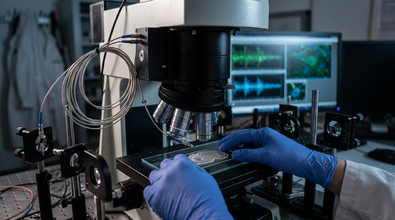
- The study addressed challenges in observing local dendritic computations, which influence neuronal signaling and plasticity, in living organisms.
- Researchers developed a 3D real-time motion correction platform for ultrafast two-photon voltage imaging of CA1 pyramidal neurons.
- The study found that bursts backpropagated more reliably than single spikes, and electro-calcium coupling decreased with increasing branch order.
- The authors concluded that dendritic structure isolates voltage from calcium signaling, potentially enabling unique intracellular pathways in distal dendrites.
- These findings enhance understanding of neuronal signal integration, which could inform future research into neurological conditions affecting dendritic function.
Unraveling the Micro-Dynamics of Neuronal Signaling
The integrity of neuronal communication is essential for all brain functions and is compromised in numerous neurological and psychiatric disorders [1, 2]. Neurons integrate information through their dendritic trees, where local electrical and biochemical events determine the cell's output. However, observing these ultrafast, fine-scale dynamics in a living brain has been a persistent technical challenge, limiting understanding of how these micro-computations contribute to neuronal activity and plasticity [3]. This gap is clinically significant, as conditions like Parkinson's disease and Alzheimer's disease involve progressive neuronal dysfunction where the underlying cellular mechanisms remain incompletely understood [4, 5]. A recent study provides new clarity by using a movement-stabilized, ultrafast two-photon voltage imaging technique to measure electrical and calcium signaling simultaneously within the dendrites of hippocampal CA1 neurons [6].
Overcoming Imaging Hurdles in Live Brains
A precise understanding of how neurons process information requires observing them at the most granular level, yet local dendritic computations have remained largely unexplored in vivo. The primary obstacle has been the difficulty of stably imaging minuscule structures at the ultrafast timescales required to capture neuronal signaling. The sub-millisecond electrical and calcium dynamics within the fine, constantly moving dendritic branches of a living animal, subject to physiological motion from respiration and circulation, have historically been too fast and unstable for conventional imaging to resolve. This has limited the ability to connect these micro-events to broader neurological function.
To address these limitations, the researchers developed a 3D real-time motion correction platform for use with ultrafast two-photon voltage imaging. This advanced microscopy uses focused infrared light to penetrate deep into living tissue with minimal scattering, enabling high-resolution visualization of electrical activity in individual neurons. The platform's critical feature is its ability to actively compensate for brain movements in real time. This stabilization ensures that the faint, rapid signals from tiny dendritic structures are captured with high fidelity, providing an unprecedented window into the fundamental processes governing neuronal communication.
Simultaneous Electrical and Calcium Measurements
Using this advanced imaging platform, the investigators focused on CA1 pyramidal neurons in the hippocampus, a cell type essential for learning and memory formation. To visualize their activity, the neurons were co-labeled with both voltage and calcium indicators. This dual-labeling strategy permitted the simultaneous measurement of two distinct but related aspects of neuronal function: the rapid changes in electrical potential and the slower, corresponding fluctuations in intracellular calcium concentration. This approach allowed the researchers to measure both somato-dendritic and electro-calcium coupling at multiple sites along a dendrite.
Measuring these two forms of coupling in tandem is essential for a complete picture of dendritic processing. Somato-dendritic coupling describes the electrical fidelity between the neuron's cell body (soma) and its dendrites, determining how effectively signals from the soma propagate outward. In parallel, electro-calcium coupling defines the relationship between an electrical event and the subsequent rise in intracellular calcium. As a critical second messenger, calcium triggers numerous downstream cellular processes, including synaptic plasticity and gene expression. By observing both signals concurrently, the study could map how electrical activity translates into biochemical change, offering deeper insight into the computational capacity of individual dendrites.
Dendritic Spike Propagation and Reliability
The study also provided a detailed characterization of how different electrical signals travel within the dendrites of CA1 neurons. The researchers analyzed isolated dendritic spikes, which are electrical impulses generated locally within the dendritic tree itself, independent of the soma. They also examined the distance-dependent backpropagation of signals originating in the soma, a process where action potentials travel backward into the dendrites. This phenomenon was assessed for naturally occurring bursts, photostimulation-evoked bursts, and single spikes, clarifying how different firing patterns influence the dendritic tree.
A central finding of this analysis was that bursts backpropagated more reliably than single spikes. This suggests that high-frequency clusters of action potentials are more effective at invading the full extent of the dendritic tree than isolated spikes are. The reliability of this backpropagation directly impacts a neuron's ability to integrate synaptic inputs and modulate synaptic strength, processes fundamental to learning. From a clinical perspective, these mechanisms are highly relevant to conditions such as epilepsy, where abnormal burst firing is a hallmark, and neurodegenerative disorders, where disruptions in dendritic signal integration can contribute to cognitive impairment.
Structural Influences on Signal Transmission
The physical architecture of a dendrite is not a passive scaffold but an active modulator of signal transmission. The study provided direct in vivo evidence for this principle, confirming that somato-dendritic coupling decreases with distance from the soma. This finding demonstrates that the electrical influence of the cell body weakens in the more remote parts of the dendritic arbor. This distance-dependent decay implies that synaptic inputs arriving closer to the soma have a more direct impact on neuronal firing, whereas inputs to distal dendrites may require stronger or more synchronized activity to influence the cell's output.
Furthermore, the investigation revealed that electro-calcium coupling decreases with increasing branch order. Dendritic branch order describes the hierarchy of branching, where smaller, higher-order branches diverge from larger, lower-order ones. This finding means the conversion of an electrical signal into a calcium signal becomes less efficient in the most distal, finely branched parts of the dendrite. This structural uncoupling of voltage from calcium signaling suggests that these distal compartments can operate with a degree of biochemical autonomy. For clinicians, understanding these structural determinants of signaling helps explain how dendritic pathology, such as the atrophy and altered branching seen in many neurodegenerative diseases, can directly disrupt neuronal computation and lead to functional deficits.
Implications for Neuronal Function and Future Research
These in vivo observations advance the model of neuronal processing, confirming that dendrites are active computational units, not just passive receivers. The study provides direct in vivo evidence for distance-dependent invasion of somatic signals into dendrites and highlights the prevalence of isolated dendritic events, underscoring the capacity for localized information processing within a single neuron. This functional compartmentalization suggests a more complex system for encoding and storing information than previously understood.
A key insight is that dendritic structure isolates voltage from calcium signaling, particularly in distal branches. This separation allows for highly localized biochemical processes that are not obligately tied to the neuron's overall electrical state, potentially enabling unique intracellular pathways in distal dendrites. Such compartmentalized signaling could facilitate complex forms of synaptic plasticity within specific dendritic segments. These fundamental findings are critical for understanding the cellular basis of disorders where dendritic health is compromised, including Alzheimer's disease, epilepsy, and certain intellectual disabilities. By clarifying the precise rules of dendritic signal integration, this research may inform future strategies aimed at modulating dendritic function to improve neuronal health.
References
1. Su J, Song Y, Zhu Z, et al. Cell–cell communication: new insights and clinical implications. Signal Transduction and Targeted Therapy. 2024. doi:10.1038/s41392-024-01888-z
2. Rasch B, Born J. About Sleep's Role in Memory. Physiological Reviews. 2013. doi:10.1152/physrev.00032.2012
3. Daneman R, Prat A. The Blood–Brain Barrier. Cold Spring Harbor Perspectives in Biology. 2015. doi:10.1101/cshperspect.a020412
4. Xu D, Yong C, Xu Y, Chen-Yu S, Peng L. Signaling pathways in Parkinson’s disease: molecular mechanisms and therapeutic interventions. Signal Transduction and Targeted Therapy. 2023. doi:10.1038/s41392-023-01353-3
5. O’Brien JT, Holmes C, Jones MS, et al. Clinical practice with anti-dementia drugs: A revised (third) consensus statement from the British Association for Psychopharmacology. Journal of Psychopharmacology. 2017. doi:10.1177/0269881116680924
6. Gonzalez KC, Terada S, Noguchi A, et al. Movement-stabilized three-dimensional optical recordings of membrane potential changes and calcium dynamics in hippocampal CA1 dendrites.. Neuron. 2026. doi:10.1016/j.neuron.2026.01.004