NMDA Receptor Ablation in Adolescent Mice Triggers Synaptic Rebound
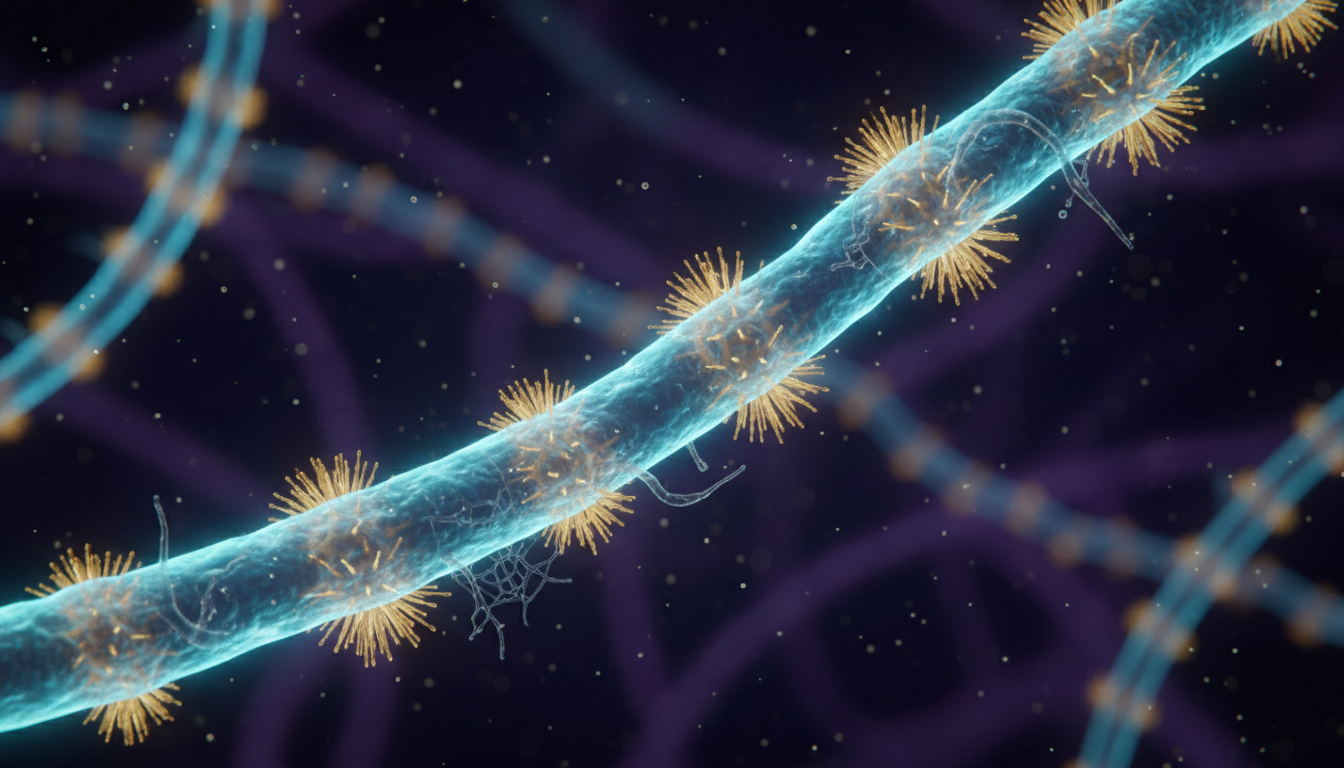
Removing NMDA receptors in the adolescent mouse prefrontal cortex causes an initial spine loss followed by a compensatory synaptic rebound.
The Clinical Lighthouse
Already a member?
or
The Clinical Lighthouse
Subscribe to read the full analysis
Full access to every article, clinical summary, and the daily evidence brief.
- Unlimited access to all articles
- Daily evidence digest
- Daily podcast with news summaries
- Lighthouse Guideline Briefings
- Ask Lumi: your guide to search, summarise, and explain the archive
- Cancel anytime